The paper demonstrates that row-column (RC) arrays have the potential to yield full three-dimensional ultrasound imaging with a greatly reduced number of elements compared to fully populated arrays. The paper describes how the various challenges for RC arrays can be overcome using synthetic aperture (SA) sequences and modified delay-and-sum beamforming to attain high quality anatomic and functional images. Resolution can approach the diffraction limit with an isotropic resolution and low side-lobe levels, and the field-of view can be expanded by using convex or lensed RC probes. Examples are shown for in-vivo volumetric B-mode images, tensor velocity imaging (TVI), and super resolution imaging. High end GPU beamforming allows for 3 orthogonal planes to be beamformed at 30 Hz, providing near real time imaging ideal for positioning the probe and improving the operator’s workflow. TVI shows the full 3-D velocity vector in a volume for revealing the full 3-D velocity vector as a function of spatial position and time for both blood velocity and tissue motion estimation. Using RC arrays with commercial contrast agents can reveal volumetric super resolution imaging with isotropic resolution in all three directions below 20 µm. RC arrays can, thus, yield full 3-D imaging at high resolution, contrast, and volumetric rates for both anatomic and functional imaging with the same number of receive channels as current commercial 1-D arrays.
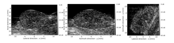 Example of volumetric row-column B-mode imaging using a Vermon 128 x 128 elements probe with a Verasonics scanner. The full volume is scanned at a frame rate of 50 Hz with an isotropic point spread function in all directions. The images shows the three orthogonal planes, but any plane in the volume can be shown.
Citation: Jensen, J. A., Schou, M., Jørgensen, L. T., Tomov, B. G., Stuart, M. B., Traberg, M. S., Taghavi, I., Øygard, S. H., Ommen, M. L., Steenberg, K., Thomsen, E. V., Panduro, N. S., Nielsen, M. B., & Sørensen, C. M. (Accepted/In press). Anatomic and Functional Imaging using Row-Column Arrays. IEEE Transactions on Ultrasonics, Ferroelectrics, and Frequency Control, https://doi.org/10.1109/TUFFC.2022.3191391
The paper can be downloaded from the CFU web-site at: DTU Orbit.
Previous Articles of the Week
The study described here investigated whether angle-independent vector flow imaging (VFI) technique estimates peak velocities in the portal vein comparably to pulsed wave Doppler (PWD).
Furthermore, intra- and inter-observer agreement was assessed in a substudy. VFI and PWD peak velocities were estimated with from intercostal and subcostal views for 32 healthy volunteers, and precision analyses were conducted.
Blinded to estimates, three physicians rescanned 10 volunteers for intra- and inter-observer agreement analyses. The precision of VFI and PWD was 18% and 28% from an intercostal view and 23% and 77% from a subcostal view, respectively.
Bias between VFI and PWD was 0.57 cm/s (p = 0.38) with an intercostal view and 9.89 cm/s (p <0.001) with a subcostal view. Intra- and inter-observer agreement was highest for VFI (inter-observer intra-class correlation coefficient: VFI 0.80, PWD 0.3; intra-observer intra-class correlation coefficient: VFI 0.90, PWD 0.69). Regardless of scan view, VFI was more precise than PWD.
This paper presents a vector flow imaging method for the integration of quantitative blood flow imaging in portable ultrasound systems.
The method combines directional transverse oscillation (TO) and synthetic aperture sequential beamforming to yield continuous velocity estimation in the whole imaging region.
Six focused emissions are used to create a high-resolution image (HRI), and a dual-stage beamforming approach is used to lower the data throughput between the probe and the processing unit. The transmit/receive focal points are laterally separated to obtain a TO in the HRI that allows for the velocity estimation along the lateral and axial directions using a phase-shift estimator.
The performance of the method was investigated with constant flow measurements in a flow rig system using the SARUS scanner and a 4.1-MHz linear array. A sequence was designed with interleaved B-mode and flow emissions to obtain continuous data acquisition. A parametric study was carried out to evaluate the effect of critical parameters.
The vessel was placed at depths from 20 to 40 mm, with beam-to-flow angles of 65°, 75°, and 90°. For the lateral velocities at 20 mm, a bias between -5% and -6.2% was obtained, and the standard deviation (SD) was between 6% and 9.6%. The axial bias was lower than 1% with an SD around 2%. The mean estimated angles were 66.70° ± 2.86°, 72.65° ± 2.48°, and 89.13° ± 0.79° for the three cases.
A proof-of-concept demonstration of the real-time processing and wireless transmission was tested in a commercial tablet obtaining a frame rate of 27 frames/s and a data rate of 14 MB/s. An in vivo measurement of a common carotid artery of a healthy volunteer was finally performed to show the potential of the method in a realistic setting. The relative SD averaged over a cardiac cycle was 4.33%.
A non-invasive method for estimating intravascular pressure changes using 2-D vector velocity is presented. The method was first validated on computational fluid dynamics (CFD) data, and with catheter measurements on phantoms.
Hereafter, the method was tested in-vivo at the carotid bifurcation and at the aortic valve of two healthy volunteers. Ultrasound measurements were performed using the experimental scanner SARUS, in combination with an 8MHz linear array transducer for experimental scans and a carotid scan, whereas a 3.5MHz phased array probe was employed for a scan of an aortic valve.
Measured 2-D fields of angle-independent vector velocities were obtained using synthetic aperture imaging. Pressure drops from simulated steady flow through six vessel geometries spanning different degrees of diameter narrowing, running from 20% – 70 %, showed relative biases from 0.35% to 12.06 %, depending on the degree of constriction. Phantom measurements were performed on a vessel with the same geometry as the 70% constricted CFD model.
The derived pressure drops were compared to pressure drops measured by a clinically used 4F catheter and to a finite element model. The proposed method showed peak systolic pressure drops of -3.0kPa±57 Pa, while the catheter and the simulation model showed -5.4kPa±52 Pa and -2.9 kPa, respectively. An in-vivo acquisition of 10 s was made at the carotid bifurcation. This produced eight cardiac cycles from where pressure gradients of -227Pa±15 Pa were found.
Lastly, the aortic valve measurement showed a peak pressure drop of -2.1 kPa over one cardiac cycle. In conclusion, pressure gradients from convective flow changes are detectable using 2-D vector velocity ultrasound.
The purpose of this work is to investigate compound lenses for row-column-addressed (RCA) ultrasound transducers for increasing the field-of-view (FOV) to a curvilinear volume region, while retaining a flat sole to avoid trapping air between the transducer sole and the patient, which would otherwise lead to unwanted reflections.
The primary motivation behind this research is to develop a RCA ultrasound transducer for abdominal or cardiac imaging, where a curvilinear volume region is a necessity. RCA transducers provide 3-D ultrasound imaging with fewer channels than fully-addressed 2-D arrays (2N instead of N2), but they have inherently limited FOV.
By increasing the RCA FOV, these transducers can be used for the same applications as fully-addressed transducers while retaining the same price range as conventional 2-D imaging due to the lower channel count. Analytical and finite element method (FEM) models were employed to evaluate design options. Composite materials were developed by loading polymers with inorganic powders to satisfy the corresponding speed of sound and specific acoustical impedance requirements. A Bi2O3 powder with a density of View the MathML source was used to decrease the speed of sound of a room temperature vulcanizing (RTV) silicone, RTV615, from View the MathML source to View the MathML source.
Using micro-balloons in RTV615 and a urethane, Hapflex 541, their speeds of sound were increased from View the MathML source to View the MathML source and from View the MathML source to View the MathML source, respectively. A diverging add-on lens was fabricated of a Bi2O3 loaded RTV615 and an unloaded Hapflex 541. The lens was tested using a RCA probe, and a FOV of 32.2° was measured from water tank tests, while the FEM model yielded 33.4°.
A wire phantom with View the MathML source diameter wires was imaged at View the MathML source down to a depth of View the MathML source using a synthetic aperture imaging sequence with single element transmissions.
The beamformed image showed that wires outside the array footprint were visible, demonstrating the increased FOV.
A non-invasive method for estimating intravascular pressure changes using 2-D vector velocity is presented.
The method was first validated on computational fluid dynamics (CFD) data, and with catheter measurements on phantoms. Hereafter, the method was tested in-vivo at the carotid bifurcation and at the aortic valve of two healthy volunteers.
Ultrasound measurements were performed using the experimental scanner SARUS, in combination with an 8MHz linear array transducer for experimental scans and a carotid scan, whereas a 3.5MHz phased array probe was employed for a scan of an aortic valve. Measured 2-D fields of angle-independent vector velocities were obtained using synthetic aperture imaging. Pressure drops from simulated steady flow through six vessel geometries spanning different degrees of diameter narrowing, running from 20% – 70 %, showed relative biases from 0.35% to 12.06 %, depending on the degree of constriction.
Phantom measurements were performed on a vessel with the same geometry as the 70% constricted CFD model. The derived pressure drops were compared to pressure drops measured by a clinically used 4F catheter and to a finite element model.
The proposed method showed peak systolic pressure drops of -3.0kPa±57 Pa, while the catheter and the simulation model showed -5.4kPa±52 Pa and -2.9 kPa, respectively. An in-vivo acquisition of 10 s was made at the carotid bifurcation. This produced eight cardiac cycles from where pressure gradients of -227Pa±15 Pa were found.
Lastly, the aortic valve measurement showed a peak pressure drop of -2.1 kPa over one cardiac cycle. In conclusion, pressure gradients from convective flow changes are detectable using 2-D vector velocity ultrasound.
Synthetic Aperture Sequential Beamforming (SASB) has shown to achieve a good resolution and high penetration depth. The low complexity at the transducer level of the beamformer makes it ideal for use with a handheld device.
SASB with a low F# (≤ 0.5) can achieve even better resolution at the cost of high grating lobes, which causes loss of contrast in the final image. In this paper, Spatial Matched Filtering (SMF) was used instead the second stage of beamformer, in an attempt to suppress the grating lobes. The advantage of SMF over SASB was investigated by pushing the limits of F#, from 1.5 to 0.5. The effect of the number of emissions used in first stage was also investigated.
A 3.3 MHz BK Ultrasound 9040 convex array was simulated in Field II on a point scatter phantom and a cyst phantom. The resolution was quantified with the full-widthhalf-max (FWHM), and the contrast was measured with the 20 dB cystic resolution. The contrast-to-noise ratio (CNR) was calculated for the cyst mimicking phantom. The results showed that SMF achieved similar resolution as SASB and improved grating lobe suppression leading to an increase in contrast.
The grating lobes caused by an F# of 0.5 are dominant in the SASB images, but not as much in SMF images. The CNR for a cyst mimicking phantom was improved 7 dB and 6 dB for SMF over SASB at depth 20 mm and 30 mm, with an F# of 0.5 and 256 emissions. The FWHM for SMF was slightly higher than SASB across all depth and parameter settings, with a maximum difference of 0.3 mm.
It was demonstrated that SMF can achieve similar resolution to SASB and for certain parameter settings improve the contrast by suppressing the grating lobe artifacts.
This Ph.D. project is based on a longstanding collaboration between physicists and engineers from the Center of Fast Ultrasound Imaging (CFU) at the Technical University of Denmark and medical doctors from the department of Radiology at Rigshospitalet. The intent of this cooperation is to validate new ultrasonic methods for future clinical use. Study I compares two B-mode ultrasound methods: the new experimental technique Synthetic Aperture Sequential Beamforming combined with Tissue Harmonic Imaging (SASB-THI), and a conventional technique combined with THI. While SASB reduces the amount of data transformation, thus enabling wireless transmission,
THI can improve resolution and image contrast, and creates fewer artifacts. Thirty-one patients with verified liver tumors were scanned and recordings with and without visible pathology were performed.
Subsequently, eight radiologists evaluated blinded to information about the technique, which B-mode images they preferred, as well as detection of pathology. Evaluation showed that the techniques were preferred equally and tumor could be detected equally well.
Study II deals with the ability of vector flow imaging (VFI) to monitor patients with arteriovenous fistulas for hemodialysis for upcoming stenosis. VFI is an angle-independent method for determining blood flow direction and velocity. Volume can be determined by integrating the velocity profile multiplied by the cross-sectional area. Nineteen patients were monitored monthly over a period of six months, and VFI estimates were compared with the reference ultrasound dilution technique (UDT). VFI volume flow values were not significantly different from UDT and had a better precision.
Concordance between VFI and UDT was high when large volume flow changes (greater than 25%) occurred between dialysis sessions. However, the methods could not be regarded as interchangeable. Study III deals with VFI’s ability to determine peak velocity in the portal vein. The commonly used ultrasound method for this is spectral Doppler, which is known to overestimate peak velocity when the angle between the blood vessel and the beam is more than 70 degrees; this overestimation becomes even larger when the angle becomes larger. VFI can determine the peak velocity angle independently.
Thirty-two healthy volunteers were scanned with spectral Doppler and VFI with two portal vein scan positions (intercostal and subcostal). The study showed that VFI estimates the same peak velocity as spectral Doppler. Furthermore, VFI has better precision and can estimate the same peak velocity with a scan position, where spectral Doppler cannot. Finally, inter- and intraobserver agreement is higher for VFI. All three studies indicate that the techniques can be used in the clinic and probably will be part of everyday practice in the near future.
To obtain accurate blood flow velocity estimates it is important to remove the clutter signal originating from tissue. Conventionally, the clutter signal has been separated from the blood signal based on the difference of their spectral frequencies. However, this approach is not enough for obtaining vector flow measurements, since the spectra overlaps at high beam-to-flow angles.
In this work a distinct approach is proposed, where the energy of the velocity spectrum is used to differentiate among the two signals. The energy based method is applied by limiting the amplitude of the velocity spectrum function to a predetermined threshold. The effect of the clutter filtering is evaluated on a plane wave (PW) scan sequence in combination with transverse oscillation (TO) and directional beamforming (DB) for velocity estimation.
The performance of the filter is assessed by comparison of the velocity estimates of the proposed filter against a conventional moving average clutter filter. The effect of tissue motion is investigated using a Field II simulation of a straight vessel with moving wall, while the direct effect of the filter on the velocity estimates is evaluated on a CFD model of a carotid bifurcation with a fixed vessel wall.
The results show that the proposed filter outperformed the moving average during moving vessel wall conditions, where standard deviations from the velocity magnitudes and angles were kept consistently below 6% and 6◦ compared to 63% and 48◦ on the moving average filter. The results on the CFD showed that on non-moving conditions the velocity estimates had minor statistical differences with errors on the magnitude of -7.95±10.1% and angles of 0.15±6.65◦ for the proposed filter compared to -5.83±9.08% and -0.12±4.48◦.
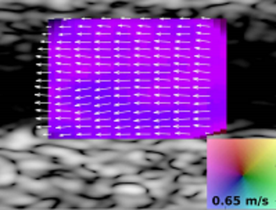
Experimental results from volumetric 3-D vector flow measurements using a 62+62 row-column addressed (RCA) array are presented.
A plane-by-plane steered transmit sequence and its post processing steps are described for obtaining 3-D vector flow in a volume. A modified version of the transverse oscillation (TO) velocity estimator is used, which exploits the focal lines generated with the tall elements of a RCA array.
Validation of the method is made in a flow-rig system where circulating blood mimicking fluid produced a steady parabolic flow profile with a flow rate of 13.7 mL/s, translating to a peak velocity of 24.1 cm/s. A volume rate of 16.4 volumes per second is obtained, and estimated flow rates based on nine steered planes within the volume are investigated.
A positive bias is found for all investigated planes lying in the range from 6.5% to 21.2% with the standard deviation being less than 4% for all cases. It is concluded that volumetric 3-D vector flow estimation is feasible with an RCA array with only 124 elements.
Portable ultrasound scanners (PUS) have, in recent years, raised a lot of attention, as they can potentially overcome some of the limitations of static scanners. However, PUS have a lot of design limitations including size and power consumption. These restrictions can compromise the image quality of the scanner. In order to overcome these restrictions, application specific integrated circuits (ASICs) are needed to implement the electronics.
In this work, a comparative study of the transmitting performance of a capacitive micromachined ultrasonic transducer (CMUT) driven by a commercial generic ultrasound transmitter and an ASIC optimized for CMUT-based PUS is presented.
A single CMUT element is pulsed with a 1% dutycycle at a frequency of 5 MHz. The DC bias voltage is 80 V and the pulsing voltage is 20 V. The acoustic performance is assessed by comparing the ultrasonic signals measured with a hydrophone both in the time and frequency domains.
The difference in normalized signal amplitude evaluated at the center frequency of the CMUT is −1.9 dB and the measured bandwidth is equivalent. The ASIC consumes only 1.3% of the total power consumption used by the commercial transmitter.
Synthetic Aperture Sequential Beamforming (SASB) has shown to achieve a good resolution and high penetration depth.
The low complexity at the transducer level of the beamformer makes it ideal for use with a handheld device. SASB with a low F# (≤ 0.5) can achieve even better resolution at the cost of high grating lobes, which causes loss of contrast in the final image. In this paper, Spatial Matched Filtering (SMF) was used instead the second stage of beamformer, in an attempt to suppress the grating lobes.
The advantage of SMF over SASB was investigated by pushing the limits of F#, from 1.5 to 0.5. The effect of the number of emissions used in first stage was also investigated. A 3.3 MHz BK Ultrasound 9040 convex array was simulated in Field II on a point scatter phantom and a cyst phantom. The resolution was quantified with the full-widthhalf-max (FWHM), and the contrast was measured with the 20 dB cystic resolution. The contrast-to-noise ratio (CNR) was calculated for the cyst mimicking phantom.
The results showed that SMF achieved similar resolution as SASB and improved grating lobe suppression leading to an increase in contrast. The grating lobes caused by an F# of 0.5 are dominant in the SASB images, but not as much in SMF images. The CNR for a cyst mimicking phantom was improved 7 dB and 6 dB for SMF over SASB at depth 20 mm and 30 mm, with an F# of 0.5 and 256 emissions. The FWHM for SMF was slightly higher than SASB across all depth and parameter settings, with a maximum difference of 0.3 mm. It was demonstrated that SMF can achieve similar resolution to SASB and for certain parameter settings improve the contrast by suppressing the grating lobe artifacts.
In this work, a 2-D vector flow imaging (VFI) method based on synthetic aperture sequential beamforming (SASB) and directional transverse oscillation is implemented on a commercially available tablet.
The SASB technique divides the beamforming process in two parts, whereby the required data rate between the probe and back-end can be reduced by a factor of 64 compared to conventional delay-and-sum focusing.
The lowered data rate enables real-time wireless transfer for both B-mode and VFI data. In the present setup, element data were acquired from a straight vessel with the SARUS research scanner and processed by a first-stage beamformer in a fixed focus. The data were subsequently transferred to an HTC Nexus 9 tablet through an ASUS RT-AC68U Wi-Fi router to simulate a wireless probe.
The second-stage beamforming of the B-mode and flow data and the velocity estimation were implemented on the tablet’s built-in GPU (Nvidia Tegra K1) through the OpenGL ES 3.1 API.
Real-time performance was achieved with rates up to 26 VFI frames per second (38 ms/frame) for concurrent processing and Wi-Fi transmission.
A row–column-addressed (RCA) 2-D array can be interpreted as two orthogonal 1-D arrays.
By transmitting with row elements and receiving the echoes through column elements or vice versa, a rectilinear volume in front of the array can be beamformed. Since the transmit and receive 1-D arrays are orthogonal to each other, only one-way focusing is possible in each transmit or receive plane.
For applications, where the scatterers are sparse, e.g., in micro-bubble tracking, this study suggests to multiply the envelope data received by the row elements when transmitting with columns as well as the data received by the column elements when transmitting with rows, to improve the focusing. In this way, at each point a two-way focused profile in both transmit and receive directions can be produced.
This paper investigates the performance of the new focusing scheme based on simulations and phantom measurements with a PZT λ/2-pitch 3 MHz 62+62 RCA 2-D transducer probe. A synthetic aperture imaging sequence with single element transmissions at a time, is designed for imaging down to 14 cm at a volume rate of 44 Hz.
In this paper, a 2-D vector flow imaging (VFI) method developed by combining synthetic aperture sequential beamforming and directional transverse oscillation is used to
image a carotid bifurcation.
Ninety-six beamformed lines are sent from the probe to the host system for each VFI frame, enabling the possibility of wireless transmission.
The velocity is estimated using a relatively inexpensive 2-D phase-shift approach, and real-time performance can be achieved in mobile devices. However, high-frame-rate velocities can be obtained by sending the data to a cluster of computers.
The objective of this study is to demonstrate the scalability of the method’s performance according to the needs of the user and the processing capabilities of the host system.
In vivo measurements of a carotid bifurcation of a 54-year-old volunteer were conducted using a linear array transducer connected to the SARUS scanner. The velocities were estimated at a rate of 134 independent frames per second (FPS) to reveal complex flow patterns.
A peak frame rate of 2140 FPS can be obtained by generating the images recursively. VFI images are shown during the systolic phase revealing the formation of a vortex in the internal carotid artery. The peak systolic velocity from a range gate in the common tract was 0.76 m s−1 with a standard deviation (SD) of 6.1%.
The mean velocity profile was calculated from the same range gate with an average SD of 7.86%.
In this study, a comparison between velocity fields for a plane wave 2-D vector flow imaging (VFI) method and a computational fluid dynamics (CFD) simulation is made.
VFI estimates are obtained from the scan of a flow phantom, which mimics the complex flow conditions in the carotid artery. Furthermore, the precision of the VFI method is investigated under laminar and complex flow conditions in vivo. The carotid bifurcation of a healthy volunteer was scanned using both fast plane wave ultrasound and magnetic resonance imaging (MRI).
The acquired MRI geometry of the bifurcation was used for fabricating an anthropomorphic flow phantom, which was also ultrasound scanned. The same geometry was used in a CFD simulation to calculate the velocity field. Results showed that similar flow patterns and vortices were estimated using CFD and VFI in the phantom.
Velocity magnitudes were estimated with a mean difference within 15 %, however, it was 23 % in the external branch. For the in vivo scan, the precision in terms of mean standard deviation (SD) of estimates aligned to the cardiac cycle was highest in the center of the common carotid artery (SD 4.7◦ for angles) and lowest in the external branch and close to the vessel wall (SD 15.0◦ for angles).
3-D Imaging using Row–Column-Addressed 2-D Arrays with a Diverging Lens: Phantom Study
A double-curved diverging lens over a flat row– column-addressed (RCA) 2-D array can extend its inherent rectilinear 3-D imaging field-of-view (FOV) to a curvilinear volume region, which is necessary for applications such as abdominal and cardiac imaging.
A concave lens with radius of 12.7 mm was manufactured using RTV664 silicone. The diverging properties of the lens were evaluated based on measurements on several phantoms. The measured 6 dB FOV in contact with a material similar to human soft tissue was less than 15% different from the theoretical predictions, i.e., a curvilinear FOV of 32°×32°.
A synthetic aperture imaging sequence with single element transmissions was designed for imaging down to 14 cm at a volume rate of 88 Hz. The performance was evaluated in terms of signal-to-noise ratio (SNR), FOV, and full-widthat-half-maximum (FWHM).
The penetration depth in a tissue mimicking phantom with 0.5 dB/(cm MHz) attenuation was 13 cm. The results of this study confirm that the proposed lens approach is an effective method for increasing the FOV, when imaging with RCA 2-D arrays.
This article describes the application of signal processing in medical ultrasound velocity estimation. Special emphasis is on the relation among acquisition methods, signal processing, and estimators employed.
The description spans from current clinical systems for one-and two-dimensional (1-D and 2-D) velocity estimation to the experimental systems for three-dimensional (3-D) estimation and advanced imaging sequences, which can yield thousands of images or volumes per second with fully quantitative flow estimates.
Here, spherical and plane wave emissions are employed to insonify the whole region of interest, and full images are reconstructed after each pulse emission for use in velocity estimation.
Current clinical ultrasound (US) systems are limited to show blood flow movement in either 1-D or 2-D.
In this paper, a method for estimating 3-D vector velocities in a plane using the transverse oscillation method, a 32×32 element matrix array, and the experimental US scanner SARUS is presented.The aim of this paper is to estimate precise flow rates and peak velocities derived from 3-D vector flow estimates.
The emission sequence provides 3-D vector flow estimates at up to 1.145 frames/s in a plane, and was used to estimate 3-D vector flow in a cross-sectional image plane. The method is validated in two phantom studies, where flow rates are measured in a flow-rig, providing a constant parabolic flow, and in a straight-vessel phantom ( ∅=8 mm) connected to a flow pump capable of generating time varying waveforms.
Flow rates are estimated to be 82.1 ± 2.8 L/min in the flow-rig compared with the expected 79.8 L/min, and to 2.68 ± 0.04 mL/stroke in the pulsating environment compared with the expected 2.57 ± 0.08 mL/stroke. Flow rates estimated in the common carotid artery of a healthy volunteer are compared with magnetic resonance imaging (MRI) measured flow rates using a 1-D through-plane velocity sequence. Mean flow rates were 333 ± 31 mL/min for the presented method and 346 ± 2 mL/min for the MRI measurements.
Several techniques can estimate the 2-D velocity vector in ultrasound. Directional beamforming (DB) estimates blood flow velocities with a higher precision and accuracy than transverse oscillation (TO), but at the cost of a high beamforming load when estimating the flow angle.
In this paper, it is proposed to use TO to estimate an initial flow angle, which is then refined in a DB step. Velocity magnitude is estimated along the flow direction using cross-correlation. It is shown that the suggested TO-DB method can improve the performance of velocity estimates compared to TO, and with a beamforming load, which is 4.6 times larger than for TO and seven times smaller than for conventional DB.
Steered plane wave transmissions are employed for high frame rate imaging, and parabolic flow with a peak velocity of 0.5 m/s is simulated in straight vessels at beamto- flow angles from 45 to 90. The TO-DB method estimates the angle with a bias and standard deviation (SD) less than 2, and the SD of the velocity magnitude is less than 2%.
When using only TO, the SD of the angle ranges from 2 to 17 and for the velocity magnitude up to 7%. Bias of the velocity magnitude is within 2% for TO and slightly larger but within 4% for TO-DB. The same trends are observed in measurements although with a slightly larger bias.
Simulations of realistic flow in a carotid bifurcation model provide visualization of complex flow, and the spread of velocity magnitude estimates is 7.1 cm/s for TO-DB, while it is 11.8 cm/s using only TO. However, velocities for TO-DB are underestimated at peak systole as indicated by a regression value of 0.97 for TO and 0.85 for TO-DB.
An in vivo scanning of the carotid bifurcation is used for vector velocity estimations using TO and TO-DB. The SD of the velocity profile over a cardiac cycle is 4.2% for TO and 3.2% for TO-DB.
Recent progress in adaptive beamforming techniques for medical ultrasound has shown that current resolution limits can be surpassed.
One method of obtaining improved lateral resolution is the Minimum Variance (MV) beamformer. The frequency domain implementation of this method effectively divides the broadband ultrasound signals into sub-bands (MVS) to conform with the narrow-band assumption of the original MV theory. This approach is investigated here using experimental Synthetic Aperture (SA) data from wire and cyst phantoms.
A 7 MHz linear array transducer is used with the SARUS experimental ultrasound scanner for the data acquisition. The lateral resolution and the contrast obtained, are evaluated and compared with those from the conventional Delay-and-Sum (DAS) beamformer and the MV temporal implementation (MVT). From the wire phantom the Full-Width-at-Half-Maximum (FWHM) measured at a depth of 52 mm, is 16.7 μm (0.08λ) for both MV methods, while the corresponding values for the DAS case are at least 24 times higher. The measured Peak-Side-lobe-Level (PSL) may reach −41 dB using the MVS approach, while the values from the DAS and MVT beamforming are above −24 dB and −33 dB, respectively. From the cyst phantom, the power ratio (PR), the contrast-to-noise ratio (CNR), and the speckle signal-to-noise ratio (sSNR) measured at a depth of 30 mm are at best similar for MVS and DAS, with values ranging between −29 dB and −30 dB, 1.94 and 2.05, and 2.16 and 2.27 respectively.
In conclusion the MVS beamformer is not suitable for imaging continuous targets, and significant resolution gains were obtained only for isolated targets.
This paper discusses methods for assessment of ultrasound image quality based on our experiences with evaluating new methods for anatomic imaging. It presents a methodology to ensure a fair assessment between competing imaging methods using clinically relevant evaluations.
The methodology is valuable in the continuing process of method optimization and guided development of new imaging methods. It includes a three phased study plan covering from initial prototype development to clinical assessment.
Recommendations to the clinical assessment protocol, software, and statistical analysis are presented. Earlier uses of the methodology has shown that it ensures validity of the assessment, as it separates the influences between developer, investigator, and assessor once a research protocol has been established. This separation reduces confounding influences on the result from the developer to properly reveal the clinical value.
The paper exemplifies the methodology using recent studies of Synthetic Aperture Sequential Beamforming tissue harmonic imaging.
Ultrasound has become highly popular to monitor atherosclerosis, by scanning the carotid artery.
The screening involves measuring the thickness of the vessel wall and diameter of the lumen. An automatic segmentation of the vessel lumen, can enable the determination of lumen diameter.
This paper presents a fully automatic segmentation algorithm, for robustly segmenting the vessel lumen in longitudinal B-mode ultrasound images.
The automatic segmentation is performed using a combination of B-mode and power Doppler images. The proposed algorithm includes a series of preprocessing steps, and performs a vessel segmentation by use of the marker-controlled watershed transform.
The ultrasound images used in the study were acquired using the bk3000 ultrasound scanner (BK Ultrasound, Herlev, Denmark) with two transducers ”8L2 Linear” and ”10L2w Wide Linear” (BK Ultrasound, Herlev, Denmark).
The algorithm was evaluated empirically and applied to a dataset of in-vivo 1770 images recorded from 8 healthy subjects. The segmentation results were compared to manual delineation performed by two experienced users.
The results showed a sensitivity and specificity of 90.41 ± 11.2 % and 97.93 ± 5.7 % (mean ± standard deviation), respectively. The amount of overlap of segmentation and manual segmentation, was measured by the Dice similarity coefficient, which was 91.25 ± 11.6 %. The empirical results demonstrated the feasibility of segmenting the vessel lumen in ultrasound scans using a fully automatic algorithm.
Constructing a double-curved row–columnaddressed (RCA) 2-D array or applying a diverging lens over the flat RCA 2-D array can extend the imaging field-of-view (FOV) to a curvilinear volume without increasing the aperture size, which is necessary for applications such as abdominal and cardiac imaging.
Extended FOV and low channel count of double-curved RCA 2-D arrays make 3-D imaging possible with equipment in the price range of conventional 2-D imaging.
This study proposes a delay-and-sum beamformation scheme specific to double-curved RCA 2-D arrays and validates its focusing ability based on simulations.
A synthetic aperture imaging sequence with single element transmissions is designed for imaging down to 14 cm at a volume rate of 88 Hz. Using a diverging lens with f-number of -1 circumscribing the underlying RCA array, the imaging quality of a double-curved λ/2-pitch 3 MHz 62+62 RCA 2-D array is investigated as a function of depth within a curvilinear FOV of 60°×60°. The simulated double-curved 2-D array exhibits the same full-width-at-halfmaximum values for a point scatterer within its curvilinear FOV at a fixed radial distance compared with a flat 2-D array within its rectilinear FOV.
The results of this study demonstrate that the proposed beamforming approach is accurate for achieving correct time-of-flight calculations, and hence avoids geometrical distortions.
Row–column-addressed CMUT arrays suffer from low receive sensitivity of the bottom elements due to a capacitive coupling to the substrate.
The capacitive coupling increases the parasitic capacitance. A simple approach to reduce the parasitic capacitance is presented, which is based on depleting the semiconductor substrate.
To reduce the parasitic capacitance by 80% the bulk doping concentration should be at most 1012 cm-3. Experimental results show that the parasitic capacitance can be reduced by 87% by applying a substrate potential of 6V relative to the bottom electrodes.
The depletion of the semiconductor substrate can be sustained for at least 10 minutes making it applicable for row–column-addressed CMUT arrays for ultrasonic imaging.
Theoretically the reduced parasitic capacitance indicates that the receive sensitivity of the bottom elements can be increased by a factor of 2:1.
Medical ultrasound has been a widely used imaging modality in healthcare platforms for examination, diagnostic purposes, and for real-time guidance during surgery. However, despite the recent advances, medical ultrasound remains the most operator-dependent imaging modality, as it heavily relies on the user adjustments on the scanner interface to optimize the scan settings.
This explains the huge interest in the subject of this PhD project entitled “AUTOMATIC ULTRASOUND SCANNING”. The key goals of the project have been to develop automated techniques to minimize the unnecessary settings on the scanners, and to improve the computer-aided diagnosis (CAD) in ultrasound by introducing new quantitative measures.
Thus, four major issues concerning automation of the medical ultrasound are addressed in this PhD project. They touch upon gain adjustments in ultrasound, automatic synthetic aperture image quality optimization, automated vessel segmentation in ultrasound, and lack of CAD in point-of-care lung ultrasound. The goals of this PhD are achieved for each of the subjects.
First, a new automated time gain compensation technique is proposed that compensates for gains of the scans in 2-D. The proposed model outperforms the current 1-D curve compensation in commercial scanners, as the 2-D topology of the scans are not fully integrated in those techniques.
Second, an automated generic technique is proposed for optimization of synthetic aperture image quality. This generic model can be used for any imaging regime using any transducer geometry.
Third, a hybrid vessel segmentation technique is proposed that combines both vector velocity estimates (VFI) and B-mode images. The technique enables the wall-to-wall visualization of VFI, as well as provides a firm ground for quantitative quantification of VFI in state-of-the-art US scanners.
Finally, a new technique is introduced to detect disease-related reverberation artifacts in lung ultrasound, thereby exploiting the full potential of this imaging modality.
The harmonic imaging mode is today a fundamental part of ultrasound imaging; it is not only used for suppressing the grating lobe artifact, but also to reduce many other acoustical artifacts in the ultrasound image.
A vital performance parameter for accepting CMUT probes as a clinical usable transducer technology is, that it can support harmonic imaging.
The large bandwidth of the CMUT is a clear advantage for harmonic imaging, but the inherent nonlinear behavior of the CMUT poses an issue as it is difficult to dissociate the harmonics generated in the tissue from the harmonic content of the transmitted signal.
This work presents how proper pulse coding of a bipolar pulser, which is present in most commercial ultrasound scanners, can reduce the intrinsic generated harmonic to fundamental pressure amplitude ratio to below −35 dB, making CMUT probes usable for clinical applications.
This paper presents a method for optimizing parameters affecting the image quality in plane wave imaging. More specifically, the number of emissions and steering angles is optimized to attain the best images with the highest frame rate possible.
The method is applied to a specific problem, where image quality for a λ-pitch transducer is compared with a λ/2-pitch transducer. Grating lobe artifacts for λ-pitch transducers degrade the contrast in plane wave images, and the impact on frame rate is studied.
Field II simulations of plane wave images are made for all combinations of the parameters, and the optimal setup is selected based on Pareto optimality. The optimal setup for a simulated 4.1-MHz λ-pitch transducer uses 61 emissions and a maximum steering angle of 20° for depths from 0 to 60 mm.
The achieved lateral full-width at half-maximum (FWHM) is 1.5λ and the contrast is −29 dB for a scatterer at 9 mm (24λ). Using a λ/2-pitch transducer and only 21 emissions within the same angle range, the image quality is improved in terms of contrast, which is −37 dB. For imaging in regions deeper than 25 mm (66λ), only 21 emissions are optimal for both the transducers, resulting in a −36 dB contrast at 34 mm (90λ). Measurements are performed using the experimental SARUS scanner connected to a λ-pitch and λ/2-pitch transducer.
A wire phantom and a tissue mimicking phantom containing anechoic cysts are scanned and show the performance using the optimized sequences for the transducers. FWHM is 1.6λ and contrast is −25 dB for a wire at 9 mm using the λ-pitch transducer. For the λ/2-pitch transducer, contrast is −29 dB.
In vivo scans of the carotid artery of a healthy volunteer show improved contrast and present fewer artifacts, when using the λ/2-pitch transducer compared with the λ-pitch. It is demonstrated with a frame rate, which is three times higher for the λ/2-pitch transducer.
Plane-Wave imaging enables very high frame rates, up to several thousand frames per second. Unfortunately the lack of transmit focusing leads to reduced image quality, both in terms of resolution and contrast.
Recently, numerous beamforming techniques have been proposed to compensate for this effect, but comparing the different methods is difficult due to the lack of appropriate tools.
PICMUS, the Plane-Wave Imaging Challenge in Medical Ultrasound aims to provide these tools.
This paper describes the PICMUS challenge, its motivation, implementation, and metrics.
In this paper, a system-level design is presented for an integrated receive circuit for a wireless ultrasound probe, which includes analog front ends and beamformation modules.
This paper focuses on the investigation of the effects of architectural design choices on the image quality. The point spread function is simulated in Field II from 10 to 160 mm using a convex array transducer. A noise analysis is performed, and the minimum signal-to-noise ratio (SNR) requirements are derived for the low-noise amplifiers (LNAs) and A/D converters (ADCs) to fulfill the design specifications of a dynamic range of 60 dB and a penetration depth of 160 mm in the B-mode image.
Six front-end implementations are compared using Nyquist-rate and modulator ADCs. The image quality is evaluated as a function of the depth in terms of lateral full-width at halfmaximum (FWHM) and −12-dB cystic resolution (CR). The designs that minimally satisfy the specifications are based on an 8-b 30-MSPS Nyquist converter and a single-bit third-order 240-MSPS modulator, with an SNR for the LNA in both cases equal to 64 dB. The mean lateral FWHM and CR are 2.4% and 7.1% lower for the architecture compared with the Nyquistrate one.
However, the results generally show minimal differences between equivalent architectures. Advantages and drawbacks are finally discussed for the two families of converters.
Radiotherapy plays an important role in modern treatment for cancer, such as cervical and prostate radiation treatment.
One of the major issue in radiotherapy is that the target should be aligned to the planned target volume prior to each treatment fraction, for which different kilovoltage (kV) and megavoltage (MV) image guided radiotherapy (IGRT) methods are developed. However, these ionization systems provide poor visualization of soft tissue, and therefore the bone matching is frequently applied as a daily tumor alignment method in cervical radiotherapy.
In this project, the Clarity 3D ultrasound system, non-invasive, non-ionizing, and good in visualization soft tissue, was used to apply uterine matching for determining the uterine shifts relative to the bone structure.
The main purpose was to investigate the reliability of the Clarity system as a possible IGRT method. We found that the conventional probe (C-probe) has limitations, while applying transabdominal US (TAUS) scan, when it came to capturing the entire uterus owing to the difficulty in probe handling.
Contrarily, the novel autoscan-probe (A-probe) was shown to be capable of capturing the entire uterus in almost all of the scans. The operators found the A-probe to be more user-friendly, and image acquisition was also performed more smoothly.
In conclusion the A-probe is a more reliable IGRT tool, and it might replace the kV- and the MV IGRT systems. In prostate radiotherapy, the movement of the prostate during radiation delivery (intrafractional prostate motion) remains challenging.
To determine the intrafractional prostate motion, various imaging techniques have been introduced, such as kV, and MV imaging, CineMRI, implanted markers and transponders. Most of the systems are based on acquiring pre- and posttreatment images, which has limitations in addressing real-time prostate motion, and includes inter-observer variations while matching image to image.
In this project, the recently developed transperineal ultrasound 4D autoscan probe is used to investigate the real-time prostate monitoring.
The purpose of this study was to investigate the feasibility of the 4D autoscan in tracking the prostate for a duration of 2 to 2.5 minutes. We found that most of the intrafractional prostate motion is less than 2 mm, which was in concordance with previously reported data. Thus, during a RapidArc/VMAT plan delivery with a beam-on time of approximately 2.5 minutes, the intrafractional prostate motion is negligible. But, since the prostate motion increases with monitoring time, the prostate displacement during 3D conformal and IMRT plans must be taken into consideration. Additionally, we conducted a prostate probe pressure study, in which TAUS scan was simulated, using a C-probe, while the prostate was continuously monitored using the TPUS autoscan.
We found that the TAUS induced pressure displacement of the prostate, in most cases, was clinically irrelevant. Since this conclusion was in opposition to most of the previously published results, which reported displacements of up to 7 mm, we discovered that 4D real-time monitoring is the most reliable method for determining the pressure displacement compared to US/US or US/CT matching methods, in which the considerable inter-observer variability, due to variations in applied probe pressure and image/image match, limits the accuracy of the readings.
In this paper, a vector flow imaging method is presented, which combines the directional transverse oscillation approach with synthetic aperture sequential beamforming to achieve an efficient estimation of the velocities.
A double oscillating field is synthesized using two sets of focused emissions separated by a distance in the lateral direction. A low resolution line (LRL) is created for each emission in the first stage beamformer, and a second beamformer provides the high resolution data used for the velocity estimation.
The method makes it possible to have continuously available data in the whole image. Therefore, high and low velocities can be estimated with a high frame rate and a low standard deviation. The first stage is a fixed-focus beamformer that can be integrated in the transducer handle, enabling the wireless transmission of the LRLs.
The approach does not require any angle compensation or prior knowledge on the beam-to-flow angle.
The feasibility of the method is demonstrated through simulations and flow rig measurements of a parabolic flow in a vessel at 90-degree beam-to-flow angle.
The mean bias obtained from 50 independent measurements is equal to -0.67% for the lateral profile and -
0.43% for the axial profile. The relative standard deviation is 3.19% and 0.47% for the lateral and axial profiles.
It is, therefore, demonstrated that vector velocity estimation can be efficiently integrated in a portable ultrasound scanner with state-of-the-art performance.
For the last decade, the field of ultrasonic vector flow imaging has gotten an increasingly attention, as the technique offers a variety of new applications for screening and diagnostics of cardiovascular pathologies.
The main purpose of this PhD project was therefore to advance the field of 3-D ultrasonic vector flow estimation and bring it a step closer to a clinical application.
A method for high frame rate 3-D vector flow estimation in a plane using the transverse oscillation method combined with a 1024 channel 2-D matrix array is presented. The proposed method is validated both through phantom studies and in vivo. Phantom measurements are compared with their corresponding reference value, whereas the in vivo measurement is validated against the current golden standard for non-invasive blood velocity estimates, based on magnetic resonance imaging (MRI). The study concludes, that a high precision was achieved and that estimates were comparable with MRI derived results.
However, the large channel count of the applied transducer hinders a commercial implementation of the 3-D method for two main reasons: The large and heavy connection
cable is impractical for clinical use, and the high channel count hampers the task of real-time processing. In a second study, some of the issue with the 2-D matrix array are solved by introducing a 2-D row-column (RC) addressing array with only 62 + 62 elements.
It is investigated both through simulations and via experimental setups in various flow conditions, if this significant reduction in the element count can still provide precise and robust 3-D vector flow estimates in a plane. The study concludes that the RC array is capable of estimating precise 3-D vector flow both in a plane and in a volume, despite the low channel count. However, some inherent new challenges are introduced with the array.
The major disadvantage with an RC transducer, is the limited field-of-view, which is restricted to the forward looking array. It is discussed, that this drawback may be solved with a diverging lens, providing a larger field-of-view, due the the dispersion of the energy. Based on the presented results it is concluded that 3-D vector flow using TO is a feasible method for obtaining angle-independent estimates of e.g. peak velocities and flow rates at a high frame rate for clinical applications.
Moreover, the RC array offers a setup allowing for real-time processing.
The paper gives a review of the current state-of-theart in ultrasound parallel acquisition systems for flow imaging using spherical and plane waves emissions.
The imaging methods are explained along with the advantages of using these very fast and sensitive velocity estimators. These experimental systems are capable of acquiring thousands of images per second for fast moving flow as well as yielding estimates of low velocity flow.
These emerging techniques allow vector flow systems to assess highly complex flow with transitory vortices and moving tissue, and they can also be used in functional ultrasound imaging for studying brain function in animals.
The paper explains the underlying acquisition and estimation methods for fast 2-D and 3-D velocity imaging and gives a number of examples. Future challenges and the potentials of parallel acquisition systems for flow imaging are also discussed.
The paper gives a review of the most important methods for blood velocity vector flow imaging (VFI) for conventional, sequential data acquisition.
This includes multibeam methods, speckle tracking, transverse oscillation, color flow mapping derived vector flow imaging, directional beamforming, and variants of these. The review covers both 2-D and 3-D velocity estimation and gives a historical perspective on the development along with a summary of various vector flow visualization algorithms.
The current state-of-the-art is explained along with an overview of clinical studies conducted and methods for presenting and using VFI. A number of examples of VFI images are presented, and the current limitations and potential solutions are discussed.
The main objective of this project was to continue the development of a synthetic aperture vector flow estimator.
This type of estimator is capable of overcoming two of the major limitations in conventional ultrasound systems: 1) the inability to scan large region of interest with high temporal resolutions; 2) the lack of capability in detecting flow other than the one along the direction of the beam.
Addressing these technical limitations would translate in the clinic as a gain in valuable clinical information and a removal of operator-dependant sources of error, which would improve the diagnosis. The main contribution of this work was the development of an angle estimator which features high accuracy and low standard deviation over the full 360◦ range.
The estimator demonstrated its capability of operating at high frame rates (> 1000 Hz), and simultaneously detecting a large range of flow velocities (0.05 – 3 m s−1 ). The estimator was also extended to a variety of geometries without major modifications, including a 2-D matrix array for full 3-D velocity estimation. Furthermore, a developed novel energy based tissue echo-canceler provided a new effective perspective for removing the tissue signal, specially when the tissue and flow spectra overlaps.
The approach was investigated with a series of flow simulations that included vessel wall movement, and demonstrated its capability of diminish the effects of a moving vessel wall in both simulations and in vivo measurements.
Finally, this thesis showed that novel information can be obtained with vector velocity methods providing quantitative estimates of blood flow and insight into the complexity of the hemodynamics dynamics. This could give the clinician a new tool in assessment and treatment of a broad range of diseases.
An efficient Fourier beamformation algorithm is presented for multistatic synthetic aperture ultrasound imaging using virtual sources (FBV).
The concept is based on the frequency domain wavenumber algorithm from radar and sonar and is extended to a multi-element transmit/receive configuration using virtual sources.
Window functions are used to extract the azimuth processing bandwidths and weight the data to reduce sidelobes in the final image. Field II simulated data and SARUS measured data are used to evaluate the results in terms of point spread function, resolution, contrast, SNR, and processing time. Lateral resolutions of 0.53 mm and 0.66 mm are obtained for FBV and DAS on point target simulated data.
Corresponding axial resolutions are 0.21 mm for FBV and 0.20 mm for DAS. The results are also consistent over different depths evaluated using a simulated phantom containing several point targets at different depths. FBV shows a better lateral resolution at all depths, and the axial and cystic resolutions of -6 dB, -12 dB and -20 dB are almost the same for FBV and DAS.
To evaluate the cyst phantom metrics, three different criteria of Power Ratio (PR), Contrast Ratio (CR), and contrast to noise ratio (CNR) have been used. Results show that the algorithms have a different performance in the cyst center and near the boundary. FBV has a better performance near the boundary, however, DAS is better in the more central area of the cyst. Measured data from phantoms are also used for evaluation. The results confirm the applicability of FBV in ultrasound and 20 times less processing time in comparison with DAS is attained.
Evaluating the results over a wide variety of parameters and having almost the same results for simulated and measured data demonstrates the ability of FBV in preserving the quality of image as DAS, while providing a more efficient algorithm with 20 times less computations.
Duplex Vector Flow Imaging (VFI) imaging is introduced as a replacement for spectral Doppler, as it automatically can yield fully quantitative flow estimates without angle correction.
Continuous VFI data over 9 s for 10 pulse cycles were acquired by a 3 MHz convex probe connected to the SARUS scanner for pulsating flow mimicking the femoral artery from a CompuFlow 1000 pump (Shelley Medical).
Data were used in four estimators based on directional transverse oscillation for velocity, flow angle, volume flow, and turbulence estimation and their respective precisions. An adaptive lag scheme gave the ability to estimate a large velocity range, or alternatively measure at two sites to find e.g. stenosis degree in a vessel.
The mean angle at the vessel center was estimated to 90.9◦±8.2◦ indicating a laminar flow from a turbulence index being close to zero (0.1 ±0.1). Volume flow was 1.29 ±0.26 mL/stroke (true: 1.15 mL/stroke, bias: 12.2%).
Measurements down to 160 mm were obtained with a relative standard deviation and bias of less than 10% for the lateral component for stationary, parabolic flow.
The method can, thus, find quantitative velocities, angles, and volume flows at sites currently inaccessible to spectral systems, and at much larger velocities and ranges than conventional systems without any angle correction making measurements less time-consuming and more correct.
Directional beamforming (DB) estimates blood flowvelocities accurately when the flow angle is known. However, forautomatically finding the flow angle a computationally expensive approach is used.
This work presents a method for estimating the flow angle using a combination of inexpensive transverse oscillation (TO) estimators and only 3 directional beamformed lines.
The suggested DB vector flow estimator is employed with steered plane wave transmissions for high frame rate imaging.Two distinct plane wave sequences are used: a short sequence(3 angles) for fast flow and an interleaved long sequence (21angles) for both slow flow and B-mode. Parabolic flow with a peak velocity of 0.5 m/s is measured at beam-to-flow angles of60◦and 90◦.
The DB method estimates the angle with a bias and standard deviation (STD) less than 2◦, and the STD of the velocity magnitude is 2.5 %. This is 7 - 8.5 % when using TO. The long sequence has a higher sensitivity, and when used forestimation of slow flow with a peak velocity of 0.04 m/s, the SDis 2.5 % and bias is 0.1 %.
This is a factor of 4 better than if the short sequence is used. The carotid bifurcation was scanned on a healthy volunteer, and the short sequence was used with TO and DB to estimate velocity vectors.
The STD of the velocity profile over a cardiac cycle was 6.1 % for TO and 4.9 % for DB.
Vector Flow Imaging (VFI) has received an increasing attention in the scientific field of ultrasound, as it enables angle independent visualization of blood flow.
VFI can be used in volume flow estimation, but a vessel segmentation is needed to make it fully automatic. A novel vessel segmentation procedure is crucial for wall-to-wall visualization, automation of adjustments, and quantification of flow in state-of-the-art ultrasound scanners. We propose and discuss a method for accurate vessel segmentation that fuses VFI data and B-mode for robustly detecting and delineating vessels.
The proposed method implements automated VFI flow measures such as peak systolic velocity (PSV) and volume flow. An evaluation of the performance of the segmentation algorithm relative to expert manual segmentation of 60 frames randomly chosen from 6 ultrasound sequences (10 frame randomly chosen from each sequence) is also presented. Dice coefficient denoting the similarity between segmentations is used for the evaluation.
The coefficient ranges between 0 and 1, where 1 indicates perfect agreement and 0 indicates no agreement. The Dice coefficient was 0.91 indicating to a very agreement between automated and manual expert segmentations.
The flowrig results also demonstrated that the PSVs measured from VFI had a mean relative error of 14.5% in comparison with the actual PSVs. The error for the PSVs measured from spectral Doppler was 29.5%, indicating that VFI is 15% more precise than spectral Doppler in PSV measurement.
3-D blood flow quantification with high spatial and temporal resolution would strongly benefit clinical research on cardiovascular pathologies.
Ultrasonic velocity techniques are known for their ability to measure blood flow with high precision at high spatial and temporal resolution. However, current volumetric ultrasonic flow methods are limited to one velocity component or restricted to a reduced field of view (FOV), e.g. fixed imaging planes, in exchange for higher temporal resolutions.
To solve these problems, a previously proposed accurate 2-D high frame rate vector flow imaging (VFI) technique is extended to estimate the 3-D velocity components inside a volume at high temporal resolutions (< 1 ms). The full 3-D vector velocities are obtained from beamformed volumetric data using synthetic aperture (SA) techniques combined with a 2-D matrix array.
The method is validated using Field II simulations of flow along a straight vessel phantom and with complex flow from a 3-D computational fluid dynamics (CFD) model of a carotid bifurcation.
Results from the simulations show that the 3-D velocity components are estimated with a mean relative bias of -12.8%, -10% and 1.42% for the Vx, Vy and Vz respectively; each presented a mean relative standard deviation of 11.8%, 12.3% and 1.11%.
Experimental 3-D vector flow estimates obtained with a 62+62 2-D row-column (RC) array with integrated apodization are presented.
A transverse oscillation (TO) velocity estimator is implemented on a 3.0 MHz RC array, to yield realtime 3-D vector flow in a cross-sectional scan plane at 750 frames per second.
The method is validated in a straight-vessel phantom (Ø = 8 mm) connected to a flow pump capable of generating timevarying carotid waveforms. The out-of-plane velocity component perpendicular to the cross section of the vessel and the crosssectional area is used to estimate volumetric flow rates. The flow rate measured from five cycles is 2.3 mL/stroke ± 0.1 mL/stroke giving a negative 9.7% bias compared to the pump settings.
It is concluded that 124 elements are sufficient to estimate 3-D vector flow, if they are positioned in a row-column wise manner.
Minimum variance beamformer (MVB) is an adaptive beamformer which provides images with higher resolution and contrast in comparison with non-adaptive beamformers like delay and sum (DAS).
It finds weight vector of beamformer by minimizing output power while keeping the desired signal unchanged.
We used the eigen-based MVB and generalized coherence factor (GCF) to further improve the quality of MVB beamformed images.
The eigen-based MVB projects the weight vector with a transformation matrix constructed from eigen-decomposing of the array covariance matrix that increases resolution and contrast. GCF is used to emphasis on coherence part of images that improves the resolution. Four different datasets provided by IUS 2016 beamforming challenge are used to evaluate the proposed method.
In comparison with DAS with rectangular weight vector, our method improved contrast about 8.52 dB and 6.20 dB for simulation and experimental contrast phantoms, respectively.
It also enhanced lateral (axial) resolution about 87% (40%) and 73% (21%) for simulated and experimental resolution phantoms, respectively.
It has been shown that row–column-addressed (RCA) 2-D arrays can be an inexpensive alternative to fully addressed 2-D arrays.
Generally imaging with an RCA 2-D array is limited to its forward-looking volume region. Constructing a double-curved RCA 2-D array or applying a diverging lens over the flat RCA 2-D array, can extend the imaging field-of-view (FOV) to a curvilinear volume without increasing the aperture size, which is necessary for applications such as abdominal and cardiac imaging.
Extended FOV and low channel count of doublecurved RCA 2-D arrays make it possible to have 3-D imaging with equipment in the price range of conventional 2-D imaging.
This study proposes a delay-and-sum (DAS) beamformation scheme specific to double-curved RCA 2-D arrays and validates its focusing ability based on simulations.
A synthetic aperture imaging (SAI) sequence with single element transmissions at a time, is designed for imaging down to 14 cm at a volume rate of 88 Hz. The curvilinear imaging performance of a λ/2-pitch 3 MHz 62+62 RCA 2-D array is investigated as a function of depth, using a diverging lens with f-number of -1.
The results of this study demonstrate that the proposed beamforming approach is accurate for achieving correct time-of-flight calculations, and hence avoids geometrical distortions.
This paper presents a novel approach for estimating 2-D flow angles using a high-frame-rate ultrasound method.
The angle estimator features high accuracy and low standard deviation (SD) over the full 360° range. The method is validated on Field II simulations and phantom measurements using the experimental ultrasound scanner SARUS and a flow rig before being tested in vivo.
An 8-MHz linear array transducer is used with defocused beam emissions. In the simulations of a spinning disk phantom, a 360° uniform behavior on the angle estimation is observed with a median angle bias of 1.01° and a median angle SD of 1.8°. Similar results are obtained on a straight vessel for both simulations and measurements, where the obtained angle biases are below 1.5° with SDs around 1°.
Estimated velocity magnitudes are also kept under 10% bias and 5% relative SD in both simulations and measurements. An in vivo measurement is performed on a carotid bifurcation of a healthy individual. A 3-s acquisition during three heart cycles is captured. A consistent and repetitive vortex is observed in the carotid bulb during systoles.
The aim of this study was to evaluate the influence of depth and underlying bone on strain ratios and shear wave speeds for three different muscles in healthy volunteers. For strain ratios the influence from different reference region-of-interest positions was also evaluated.
Material and methods: Ten healthy volunteers (five males and five females) had their biceps brachii, gastrocnemius, and quadriceps muscle examined with strain- and shear wave elastography at three different depths and in regions located above bone and beside bone. Strain ratios were averaged from cine-loops of 10 s length, and shear wave speeds were measured 10 times at each target point.
The distance from the skin surface to the centre of each region-of-interest was measured. Measurements were evaluated with descriptive statistics and linear regression.
Results: Linear regression showed a significant influence on strain ratio measurements from the reference region-of-interest position, i.e. being above the same structures as the target region-of-interest or not (means: 1.65 and 0.78; (P < 0.001)). For shear wave speeds, there was a significant influence from depth and location above or beside bone (P = 0.011 and P = 0.031).
Conclusion: Strain ratio values depend significantly on reference and target region-of-interest being above the same tissue, for instance bone. Strain ratios were not influenced by depth in this study. Shear wave speeds decreased with increasing scanning depth and if there was bone below the region-of-interest.
A signal based algorithm resulting in increased depth resolution is presented for medical ultrasound. It relies on multiple foci beamforming that is enabled by current ultrasound imaging systems.
The concept stems from optical microscopy and is translated here into ultrasound using the Field II simulation software.
A 7 MHz linear transducer is used to scan a single point scatterer phantom that can move in the axial direction. Individual beamformer outputs from 3 different foci are post-processed using the highly-dependent on focusing errors, metric of sharpness to estimate the position of the point scatter.
A 37.8 μm uncertainty in depth estimation is achieved, which attains an almost 3-fold improvement compared to conventional ultrasound imaging axial resolution.
Future work on the development of this algorithm requires experimental validation in tissue-like materials that provide strong aberrations.
Synthetic Aperture (SA) imaging produces high-quality images and velocity estimates of both slow and fast flow at high frame rates. However, grating lobe artifacts can appear both in transmission and reception. These affect the image quality and the frame rate.
Therefore optimization of parameters effecting the image quality of SA is of great importance, and this paper proposes an advanced procedure for optimizing the parameters essential for acquiring an optimal image quality, while generating high resolution SA images.
Optimization of the image quality is mainly performed based on measures such as F-number, number of emissions and the aperture size. They are considered to be the most contributing acquisition factors in the quality of the high resolution images in SA.
Therefore, the performance of image quality is quantified in terms of full-width at half maximum (FWHM) and the cystic resolution (CTR). The results of the study showed that SA imaging with only 32 emissions and maximum sweep angle of 22 degrees yields a very good image quality compared with using 256 emissions and the full aperture size.
Therefore the number of emissions and the maximum sweep angle in the SA can be optimized to reach a reasonably good performance, and to increase the frame rate by lowering the required number of emissions.
All the measurements are performed using the experimental SARUS scanner connected to a λ/2-pitch transducer. A wire phantom and a tissue mimicking phantom containing anechoic cysts are scanned using the optimized parameters for the transducer.
Measurements coincide with simulations.
A framework for simulating ultrasound imaging based on first order nonlinear pressure–velocity relations
An ultrasound imaging framework modeled with the first order nonlinear pressure–velocity relations (NPVR) based simulation and implemented by a half-time staggered solution and pseudospectral method is presented in this paper.
The framework is capable of simulating linear and nonlinear ultrasound propagation and reflections in a heterogeneous medium with different sound speeds and densities.
It can be initialized with arbitrary focus, excitation and apodization for multiple individual channels in both 2D and 3D spatial fields.
The simulated channel data can be generated using this framework, and ultrasound image can be obtained by beamforming the simulated channel data. Various results simulated by different algorithms are illustrated for comparisons.
The root mean square (RMS) errors for each compared pulses are calculated. The linear propagation is validated by an angular spectrum approach (ASA) with a RMS error of 3% at the focal point for a 2D field, and Field II with RMS errors of 0.8% and 1.5% at the electronic and the elevation focuses for 3D fields, respectively.
The accuracy for the NPVR based nonlinear propagation is investigated by comparing with the Abersim simulation for pulsed fields and with the nonlinear ASA for monochromatic fields.
The RMS errors of the nonlinear pulses calculated by the NPVR and Abersim are respectively 2.4%, 7.4%, 17.6% and 36.6% corresponding to initial pressure amplitudes of 50 kPa, 200 kPa, 500 kPa and 1 MPa at the transducer.
By increasing the sampling frequency for the strong nonlinearity, the RMS error for 1 MPa initial pressure amplitude is reduced from 36.6% to 27.3%.
This work presents the first in vivo results of 2-D high frame rate vector velocity imaging for transthoracic cardiac imaging.
Measurements are made on a healthy volunteer using the SARUS experimental ultrasound scanner connected to an intercostal phased-array probe.
Two parasternal long-axis view (PLAX) are obtained, one centred at the aortic valve and another centred at the left ventricle. The acquisition sequence was composed of 3 diverging waves for high frame rate synthetic aperture flow imaging.
For verification a phantom measurement is performed on a transverse straight 5 mm diameter vessel at a depth of 100 mm in a tissue-mimicking phantom. A flow pump produced a 2 ml/s constant flow with a peak velocity of 0.2 m/s.
The average estimated flow anglein the ROI was 86.22◦ ± 6.66◦ with a true flow angle of 90◦. A relative velocity bias of −39% with a standard deviation of 13% was found. In-vivo acquisitions show complex flow patterns in the heart.
In the aortic valve view, blood is seen exiting the left ventricle cavity through the aortic valve into the aorta during the systolic phase of the cardiac cycle. In the left ventricle view, blood flow is seen entering the left ventricle cavity through the mitral valve and splitting in two ways when approximating the left ventricle wall.
The work presents 2-D velocity estimates on the heart from a non-invasive transthoracic scan. The ability of the method detecting flow regardless of the beam angle could potentially reveal a more complete view of the flow patterns presented on the heart.
This paper presents an in-house developed 2-D capacitive micro machined ultrasonic transducer (CMUT) applied for 3-D blood flow estimation.
The probe breaks with conventional transducers in two ways; first, the ultrasonic pressure field is generated from thousands of small vibrating micro machined cells, and second, elements are accessed by row and/or column indices.
The 62+62 2-D row-column addressed prototype CMUT probe was used for vector flow estimation by transmitting focused ultrasound into a flow-rig with a fully developed parabolic flow. The beam-to-flow angle was 90◦. The received data was beamformed and processed offline.
A transverse oscillation (TO) velocity estimator was used to estimate the 3-D vector flow along a line originating from the center of the transducer.
The estimated velocities in the lateral and axial direction were close to zero as expected.In the transverse direction a characteristic parabolic velocity profile was estimated with a peak velocity of 0.48m/s ± 0.02 m/s in reference to the expected 0.54 m/s. The results presented are the first 3-D vector flow estimates obtained with a row-column CMUT probe, which demonstrates that the CMUT technology is feasible for 3-D flow estimation.
This paper presents a novel beamformer architecture for a low-cost receiver front-end, and investigates if the image quality can be maintained.
The system is oriented to the development of a hand-held wireless ultrasound probe based on Synthetic Aperture Sequential Beamforming, and has the advantage of effectively reducing circuit complexity and power dissipation.
The array of transducers is divided into sub-apertures, in which the signals from the single channels are aligned through a network of cascaded gradient delays, and summed in the analog domain before A/D conversion. The delay values are quantized to simplify the shifting unit, and a single A/D converter is needed for each sub-aperture yielding a compact, low-power architecture that can be integrated in a single chip.
A simulation study was performed using a 3.75 MHz convex array, and the point spread function (PSF) for different configurations was evaluated in terms of lateral full-width-at-half-maximum (FWHM) and −20 dB cystic resolution (CR). Several setups were simulated varying the sub-aperture size N and the quantization step, and design constraints were obtained comparing the PSF to that of an ideal non-quantized system.
The PSF is shown for N = 32 with a quantization step of 12 ns. For this configuration, the FWHM is degraded by 0.25% and the CR is 8.70% lower compared to the ideal situation. The results demonstrate that the gradient beamformer provides an adequate image quality, and open the way to a fully-integrated chip for a compact, low-cost, wireless ultrasound probe.
Chronic venous disease is a common condition leading to varicose veins, leg edema, post-thrombotic syndrome and venous ulcerations.
Ultrasound (US) is the main modality for examination of venous disease. Color Doppler and occasionally spectral Doppler US (SDUS) are used for evaluation of the venous flow. Peak velocities measured by SDUS are rarely used in a clinical setting for evaluating chronic venous disease due to inadequate reproducibility mainly caused by the angle dependency of the estimate.
However, estimations of blood velocities are of importance in characterizing venous disease. Transverse Oscillation US (TOUS), a non-invasive angle independent method, has been implemented on a commercial scanner.
TOUS’s advantage compared to SDUS is a more elaborate visualization of complex flow. The aim of this study was to evaluate, whether TOUS perform equal to SDUS for recording velocities in the veins of the lower limbs.
Four volunteers were recruited for the study. A standardized flow was provoked with a cuff compression-decompression system placed around the lower leg. The average peak velocity in the popliteal vein of the four volunteers was 151.5 cm/s for SDUS and 105.9 cm/s for TOUS (p <0.001). The average of the peak velocity standard deviations (SD) were 17.0 cm/s for SDUS and 13.1 cm/s for TOUS (p <0.005).
The study indicates that TOUS estimates lower peak velocity with improved SD when compared to SDUS. TOUS may be a tool for evaluation of venous disease providing quantitative measures for the evaluation of venous blood flow.
The main purpose of this PhD project was to develop an ultrasonic method capable of determining intravascular pressure changes non-invasively.
Measuring pressure variations is used clinically as a diagnostic marker for the physiological state of a cardiovascular region.
Current clinical procedures for assessing pressure changes are by means of invasive devices such as pressure sensing catheters. Such devices suffer severe limitations as they are invasive and require the use of ionizing radiation for guidance and positioning. To overcome the concerns related to the use of invasive pressure catheters this project introduces a method that derives pressure changes from 2-D vector velocity flow data acquired non-invasively.
The method is based on the Navier-Stokes equations and is tested on fabricated flow models. Results from the flow models are compared with simulations from finite element modeling. The developed technique showed a standard deviation and bias across constricted flow domains of 9 % and 8 %, respectively. Finally, the first in-vivo examples of deriving pressure changes from 2-D vector velocity ultrasound data is presented.
Based on the presented results it is concluded that non-invasive determination of pressure changes from 2-D flow data is feasible. However, when transferring the method into clinical practice, where blood vessels follow more complex flow geometries, the influence of out-of-plane flow movement becomes increasingly more important.
Therefore, for scans using a 1-D transducer it is crucial that the out-of-plane flow component is negligible.
This paper presents a novel automatic method for detection of B-lines (comet-tail artifacts) in lung ultrasound scans.
B-lines are the most commonly used artifacts for analyzing the pulmonary edema. They appear as laser-like vertical beams, which arise from the pleural line and spread down without fading to the edge of the screen. An increase in their number is associated with presence of edema.
All the scans used in this study were acquired using a BK3000 ultrasound scanner (BK Ultrasound, Denmark) driving a 192-element 5.5 MHz wide linear transducer (10L2W, BK Ultrasound).
The dynamic received focus technique was employed to generate the sequences. Six subjects, among those three patients after major surgery and three normal subjects, were scanned once and Six ultrasound sequences each containing 50 frames were acquired. The proposed algorithm was applied to all 300 in-vivo lung ultrasound images.
The pleural line is first segmented on each image and then the B-line artifacts spreading down from the pleural line are detected and overlayed on the image. The resulting 300 images showed that the mean lateral distance between B-lines detected on images acquired from patients decreased by 20% in compare with that of normal subjects.
Therefore, the method can be used as the basis of a method of automatically and qualitatively characterizing the distribution of B-lines.
Safety Assessment of Advanced Imaging Sequences II: Simulations
An automatic approach for simulating the emitted pressure, intensity, and MI of advanced ultrasound imaging sequences is presented.
It is based on a linear simulation of pressure fields using Field II, and it is hypothesized that linear simulation can attain the needed accuracy for predicting Mechanical Index (MI) and Ispta.3 as required by FDA.
The method is performed on four different imaging schemes and compared to measurements conducted using the SARUS experimental scanner. The sequences include focused emissions with an F-number of 2 with 64 elements that generate highly non-linear fields.
The simulation time is between 0.67 ms to 2.8 ms per emission and imaging point, making it possible to simulate even complex emission sequences in less than 1 s for a single spatial position. The linear simulations yield a relative accuracy on MI between -12.1% to 52.3% and for Ispta.3 between -38.6% to 62.6%, when using the impulse response of the probe estimated from an independent measurement.
The accuracy is increased to between -22% to 24.5% for MI and between -33.2% to 27.0% for Ispta.3, when using the pressure response measured at a single point to scale the simulation. The spatial distribution of MI and Ita.3 closely matches that for the measurement, and simulations can therefore be used to select the region for measuring the intensities, resulting in a significant reduction in measurement time. It can validate emission sequences by showing symmetry of emitted pressure fields, focal position, and intensity distribution.
A method for rapid measurement of intensities (Ispta), mechanical index (MI), and probe surface temperature for any ultrasound scanning sequence is presented.
It uses the scanner’s sampling capability to give an accurate measurement of the whole imaging sequence for all emissions to yield the true distributions.
The method is several orders of magnitude faster than approaches using an oscilloscope, and it also facilitates validating the emitted pressure field and the scanner’s emission sequence software.
It has been implemented using the experimental SARUS scanner and the Onda AIMS III intensity measurement system (Onda Corporation, Sunnyvale, CA, USA). Four different sequences have been measured: a fixed focus emission, a duplex sequence containing B-mode and flow emissions, a vector flow sequence with B-mode and flow emissions in 17 directions, and finally a synthetic aperture (SA) duplex flow sequence. A BK8820e (BK Medical, Herlev, Denmark) convex array probe is used for the first three sequences and a BK8670 linear array probe for the SA sequence.
The method is shown to give the same intensity values within 0.24% of the AIMS III Soniq 5.0 (Onda Corporation, Sunnyvale, CA, USA) commercial intensity measurement program.
The approach can measure and store data for a full imaging sequence in 3.8 to 8.2 s per spatial position. Based on Ispta, MI, and probe surface temperature, the method gives the ability to determine whether a sequence is within US FDA limits, or alternatively indicate how to scale it to be within limits.
The synthetic aperture (SA) technique can be used for achieving real-time volumetric ultrasound imaging using 2-D row-column addressed transducers.
This paper investigates SA volumetric imaging performance of an in-house prototyped 3 MHz λ/2-pitch 62+62 element piezoelectric 2-D row-column addressed transducer array. Utilizing single element transmit events, a volume rate of 90 Hz down to 14 cm deep is achieved.
Data are obtained using the experimental ultrasound scanner SARUS with a 70 MHz sampling frequency and beamformed using a delay-and-sum (DAS) approach. A signal-to-noise ratio of up to 32 dB is measured on the beamformed images of a tissue mimicking phantom with attenuation of 0.5 dB cm−1 MHz−1, from the surface of the probe to the penetration depth of 300λ. Measured lateral resolution as Full-Width-at-Half-Maximum (FWHM) is between 4λ and 10λ for 18 % to 65 % of the penetration depth from the surface of the probe.
The averaged contrast is 13 dB for the same range. The imaging performance assessment results may represent a reference guide for possible applications of such an array in different medical fields.
For 3-D ultrasound imaging with row-column addressed 2-D arrays, the two orthogonal 1-D transmit and receive arrays are both used for one-way focusing in the lateral and elevation directions separately and since they are not in the same plane, the two-way focusing is the same as one-way focusing.
However, the achievable spatial resolution and contrast of the B-mode images in Delay and Sum (DAS) beamforming are limited by the aperture size and by the operating frequency.
This paper, investigates Spatial Matched Filter (SMF) beamforming on row-column addressed 2-D arrays to increase spatial resolution.
The performance is investigated on both simulated and experimentally collected 3-D data by comparing the Point Spread Functions (PSFs) and the phantom images obtained with standard DAS and with SMF.
Results show that the SMF beamformer outperforms DAS in both simulated and experimental trials and that a higher contrast resolution can be achieved by SMF beamforming (i.e., narrower main lobe and lower side lobes).
The 6dB, 20dB and 40dB cystic resolution for a DAS simulated PSF at (0,0,30)mm are 1.22mm, 3.54mm and 7.46mm, for SMF beamforming they are 1.11mm, 2.33mm and 5.42mm accordingly. For measured RF-data of an iron needle facing toward the transducer positioned at (0,0,32.5)mm along the central axis, the 6dB, 20dB and 40dB cystic resolution for DAS beamforming are 1.99mm, 2.19mm and 4.26mm, and they are 0.8mm, 2.06mm and 4.18mm for SMF beamforming accordingly.
SMF beamforming increases the contrast resolution which turns into a better quality of the B-mode images.
TO measurements were performed with a 3 MHz convex probe (BK medical 8820e, Herlev, Denmark) connected to the experimental ultrasound scanner SARUS (Synthetic Aperture Real-time Ultrasound Scanner). SDU velocity measurements were performed with a commercial ultrasound scanner (BK 3000, BK Ultrasound, Herlev Denmark) and a convex probe (BK ultrasound 6C2, Herlev, Denmark).
Ten healthy volunteers were scanned, and recordings of the portal flow during 3-5 heartbeats were conducted with an intercostal and subcostal view. Intercostal TO peak velocities were not significantly different from SDU peak velocities (TO=0.203m/s, SDU=0.202m/s, p=0.94). Subcostal and Intercostal obtained TO values were not significantly different (intercostal mean TO=0.203m/s, subcostal mean TO=0.180m/s, p=0.26).
SDU values obtained intercostal and subcostal were significantly different (intercostal mean SDU=0.202m/s, subcostal mean SDU=0.320m/s, p<0.001). Standard deviation for TO beam-to-flow angle was 10.3°- 91.5°, indicating a large beam-to-flow angle variability in the portal vein. This can affect the peak velocity estimation, and is not addressed in SDU.
The TO convex array implementation provides the first vector velocity measurements below 60mm (mean 89mm), and is a useful alternative for flow estimation in abdominal ultrasound. It may provide new information of abdominal fluid dynamics and yield both velocity and angle estimates for a more realistic flow characterization.
In the ascending aorta, atherosclerotic plaque formation, which is a risk factor for cerebrovascular events, most often occurs along the inner curvature.
Atherosclerosis is a multifactorial disease, but the predilection site for the aortic vessel degradation is probably flow dependent.
To better understand the aortic flow and especially the complex flow patterns, the ascending aorta was scanned intraoperatively in patients undergoing heart surgery using the angle-independent vector velocity ultrasound method Transverse Oscillation (TO).
The primary aim of the study was to analyze systolic backflow in relation to atherosclerosis. Thirteen patients with normal aortic valves were included in to the study.
TO implemented on a conventional US scanner (ProFocus 2202 UltraView, BK Medical, Herlev, Denmark) with a linear array transducer (8670, BK Medical, Herlev, Denmark) was used intraoperatively on the ascending aorta in long axis view.
The presence of systolic backflow, visualized with TO, was correlated to aortic atherosclerosis, to systolic velocities obtained with transesophageal echocardiography and cardiac output obtained with pulmonary artery catheter thermodilution, to gender, age, aortic diameter, left ventricular ejection fraction (LVEF) and previous myocardial infarctions (MI). Systolic backflow in the ascending aorta was present for 38% (n=5) of the patients.
The location of the backflow was strongly associated to the location of the plaques (p<0.005), and backflow was associated to high systolic velocities (p<0.05). The other obtained parameters were not associated to systolic backflow. It was shown that systolic backflow is a common flow feature in the ascending aorta, and that backflow is associated to atherosclerotic plaques and systolic velocities.
The study indicates that vector flow imaging using TO can provide important blood flow information in the assessment of atherosclerosis.
In-Vivo High Dynamic Range Vector Flow Imaging
Current vector flow systems are limited in their detectable range of blood flow velocities.
Previous work on phantoms has shown that the velocity range can be extended using synthetic aperture directional beamforming combined with an adaptive multi-lag approach.
This paper presents a first in-vivo example with a high dynamic velocity range. Velocities with an order of magnitude apart are detected on the femoral artery of a 41 years old healthy individual.
Three distinct heart cycles are captured during a 3 secs acquisition. The estimated vector velocities are compared with each other within the heart cycle. The relative standard deviation of the measured velocity magnitude between the three peak systoles was found to be 5.11% with a standard deviation on the detected angle of 1.06◦ . In the diastole, it was 1.46% and 6.18◦ , respectively.
Results prove that the method is able to estimate flow in-vivo and provide quantitative results in a high dynamic velocity range. Providing velocity measurements during the whole cardiac cycle for both arteries and veins.
Clinical applications of plane wave imaging necessitate the creation of high-quality images with the highest possible frame rate for improved blood flow tracking and anatomical imaging.
However, linear array transducers create grating lobe artefacts, which degrade the image quality especially in the near field for λ-pitch transducers.
Artefacts can only partly be suppressed by increasing the number of emissions, and this paper demonstrates how the frame rate can be increased without loss of image quality by using λ/2-pitch transducers.
The number of emissions and steering angles are optimized in a simulation study to get the best images with as high a frame rate as possible. The optimal setup for a simulated 4.1 MHz λ-pitch transducer is 73 emissions and a maximum steering of 22◦ .
The achieved FWHM is 1.3λ and the cystic resolution is -25 dB for a scatter at 9 mm. Only 37 emissions are necessary within the same angle range when using a λ/2-pitch transducer, and the cystic resolution is reduced to -56 dB. Measurements are performed with the experimental SARUS scanner connected to a λ-pitch and λ/2-pitch transducer.
A wire phantom and a tissue mimicking phantom containing anechoic cysts are scanned and show the performance using the optimized sequences for the transducers.
Measurements confirm results from simulations, and the λ-pitch transducer show artefacts at undesirable strengths of -25 dB for a low number of emissions.
In this study, a method for estimating 3-D vector velocities at very high frame rate using continuous data acquisition is presented.
An emission sequence was designed to acquire real-time continuous data in one plane. The transverse oscillation (TO) method was used to estimate 3-D vector flow in a carotid flow phantom and in vivo in the common carotid artery of a healthy 27-year old female.
Based on the out-of-plane velocity component during four periodic cycles, estimated flow rates in an experimental setup was 2.96 ml/s ± 0.35 ml/s compared to the expected 3.06 ml/s ± 0.09 ml/s. In the in vivo measurements, three heart cycles acquired at 2.1 kHz showed peak out-of-plane velocities of 83 cm/s, 87 cm/s and 90 cm/s in agreement with the 92 cm/s found with spectral Doppler.
Mean flow rate was estimated to 257 ml/min. The results demonstrate that accurate real-time 3- D vector velocities can be obtained using the TO method, which can be used to improve operator-independece when examining blood flow in vivo, thereby increasing accuracy and consistency.
A method for estimating vector velocities using transverse oscillation (TO) combined with directional beamforming is presented.
Directional Transverse Oscillation (DTO) is selfcalibrating, which increase the estimation accuracy and finds the lateral oscillation period automatically.
A normal focused field is emitted and the received signals are beamformed in the lateral direction transverse to the ultrasound beam. A lateral oscillation is obtained by having a receive apodization waveform with two separate peaks. The IQ data are obtained by making a Hilbert transform of the directional signal, and a modified TO estimator can be used to find both the lateral and axial velocity.
The approach is self-calibrating as the lateral oscillation period directly is estimated from the directional signal through a Fourier transform. The approach was implemented on the SARUS scanner using a BK Medical 8820e transducer with a focal point at 105.6 mm (F#=5) for Vector Flow Imaging (VFI). A 6 mm radius tube in a circulating flow rig was scanned and the parabolic volume flow of 112.7 l/h (peak velocity 0.55 m/s) measured by a Danfoss Magnetic flow meter for reference.
Velocity estimates for DTO are found for 32 emissions at a 90 degrees beam-to-flow angle at a vessel depth of 30 mm. The standard deviation (SD) drops from 9.14% for TO to 5.4%, when using DTO. The bias is -5.05% and the angle is found within +/- 3.93 degrees. At 70 mm a relative SD of 7% is obtained, the bias is -1.74%, and the angle is found within +/- 2.6 degrees showing a low bias across depths.
This paper presents a method for estimating 2-D vector velocities using plane waves and transverse oscillation.
The approach uses emission of a low number of steered plane waves, which result in a high frame rate and continuous acquisition of data for the whole image.
A transverse oscillating field is obtained by filtering the beamformed RF images in the Fourier domain using a Gaussian filter centered at a desired oscillation frequency. Performance of the method is quantified through measurements with the experimental scanner SARUS and the BK 2L8 linear array transducer. Constant parabolic flow in a flow rig phantom is scanned at beam-to-flow angles of 90, 75, and 60◦ .
The relative bias is between -1.4 % and -5.8 % and the relative std. between 5 % and 8.2 % for the lateral velocity component at the measured beam-to-flow angles. The estimated flow angle is 73.4◦± 3.6◦ for the measurement at 75◦ .
Measurement of pulsatile flow through a constricted vessel demonstrate the application of the method in a realistic flow environment with large spatial and temporal flow gradients.
The concept of 2-D row-column (RC) addressed arrays for 3-D imaging have shown to be an interesting alternative to 2-D matrix array, due to the reduced channel count. However, the properties for RC arrays to estimate blood velocities have never been reported, which is of great importance for a clinical implementation of this type of array.
The aim of this study is, thus, to develop a technique for estimating 3-D vector flow with a RC array using the transverse oscillation (TO) method.
The properties are explored both in a simulation study and with a prototype probe for experimental use. In both setups, a 124 channel 2-D RC array with integrated apodization, pitch = 270 µm and a center frequency of 3.0 MHz was used.
The performance of the estimator was tested on a simulated vessel (Ø = 12 mm) with a parabolic flow profile and a peak velocity of 1 m/s. Measurements were made in a flowrig (Ø = 12 mm) containing a laminar parabolic flow and a peak velocity of 0.54 m/s. Data was sampled and stored on the experimental ultrasound scanner SARUS. Simulations yields relative mean biases at (- 1.1%, -1.5%, -1.0%) with mean standard deviations of σ˜ were (8.5%, 9.0%, 1.4%) % for (vx, vy, vz) from a 3-D velocity vector in a 15◦ rotated vessel with a 75◦ beam-to-flow angle. In the experimental setup with a 90◦ beam-to-flow angle, the relative mean biases were (-2.6%, -1.3% , 1.4%) with a relative standard deviation of (5.0%, 5.2%, 1.0%) for the respective transverse, lateral and axial velocity component.
Synthetic aperture (SA) imaging can be used to achieve real-time volumetric ultrasound imaging using 2-D array transducers. The sensitivity of SA imaging is improved by maximizing the acoustic output, but one must consider the limitations of an ultrasound system, both technical and biological.
This paper investigates the in vivo applicability and sensitivity of volumetric SA imaging. Utilizing the transmit events to generate a set of virtual point sources, a frame rate of 25 Hz for a 90° × 90° field-of-view was achieved. data were obtained using a 3.5 MHz 32 × 32 elements 2-D phased array transducer connected to the experimental scanner (SARUS).
Proper scaling is applied to the excitation signal such that intensity levels are in compliance with the U.S. Food and Drug Administration regulations for in vivo ultrasound imaging. The measured Mechanical Index and spatial-peak-temporal-average intensity for parallel beam-forming (PB) are 0.83 and 377.5mW/cm2, and for SA are 0.48 and 329.5mW/cm2. A human kidney was volumetrically imaged with SA and PB techniques simultaneously.
Two radiologists for evaluation of the volumetric SA were consulted by means of a questionnaire on the level of details perceivable in the beam-formed images. The comparison was against PB based on the in vivo data.
The feedback from the domain experts indicates that volumetric SA images internal body structures with a better contrast resolution compared to PB at all positions in the entire imaged volume.
Furthermore, the autocovariance of a homogeneous area in the in vivo SA data, had 23.5% smaller width at the half of its maximum value compared to PB. © (2015) COPYRIGHT Society of Photo-Optical Instrumentation Engineers (SPIE).
Downloading of the abstract is permitted for personal use only.
Time gain compensation (TGC) is essential to ensure the optimal image quality of the clinical ultrasound scans. When large fluid collections are present within the scan plane, the attenuation distribution is changed drastically and TGC compensation becomes challenging.
This paper presents an automated hierarchical TGC (AHTGC) algorithm that accurately adapts to the large attenuation variation between different types of tissues and structures.
The algorithm relies on estimates of tissue attenuation, scattering strength, and noise level to gain a more quantitative understanding of the underlying tissue and the ultrasound signal strength. The proposed algorithm was applied to a set of 44 in vivo abdominal movie sequences each containing 15 frames. Matching pairs of in vivo sequences, unprocessed and processed with the proposed AHTGC were visualized side by side and evaluated by two radiologists in terms of image quality.
Wilcoxon signed-rank test was used to evaluate whether radiologists preferred the processed sequences or the unprocessed data. The results indicate that the average visual analogue scale (VAS) is positive ( p-value: 2.34 × 10⁻¹³) and estimated to be 1.01 (95% CI: 0.85; 1.16) favoring the processed data with the proposed AHTGC algorithm.
© (2015) COPYRIGHT Society of Photo-Optical Instrumentation Engineers (SPIE). Downloading of the abstract is permitted for personal use only.
This study investigates the planarity of an assumed planar surface made of an elasomer fixed on its perimeter by a square acrylic frame.
The central part of this surface is insonified with two different linear array transducers (types 8811 and 8670, BK Medical), yielding 11 images forming two 3D data sets. The change in flatness of the surface was determined by cross-correlation of the matrix of received signals.
The cross-correlation was calculated between signals from the same image and as well as between signals that belonged to different images. The maximal change over the entire surface investigated is found to be in the order of a wavelength at about 12 MHz.
Specifically, the surface showed weak concave bending for the two different transducers. The data was validated using the cross-correlation coefficient function.
This paper investigates the effect of transducerintegrated apodization in row–column-addressed arrays and presents a beamforming approach specific for such arrays.
Row–column addressing 2-D arrays greatly reduces the number of active channels needed to acquire a 3-D volume. A disadvantage of row–column-addressed arrays is an apparent ghost effect in the point spread function caused by edge waves.
This paper investigates the origin of the edge waves and the effect of introducing an integrated apodization to reduce the ghost echoes. The performance of a λ/2-pitch 5-MHz 128 + 128 row–column-addressed array with different apodizations is simulated.
A Hann apodization is shown to decrease imaging performance away from the center axis of the array because of a decrease in main lobe amplitude. Instead, a static roll-off apodization region located at the ends of the line elements is proposed. In simulations, the peak ghost echo intensity of a scatterer at (x,y, z) = (8, 3, 30) mm was decreased by 43 dB by integrating roll-off apodization into the array.
The main lobe was unaffected by the apodization. Simulations of a 3-mm-diameter anechoic blood vessel at 30 mm depth showed that applying the transducer-integrated apodization increased the apparent diameter of the vessel from 2.0 mm to 2.4 mm, corresponding to an increase from 67% to 80% of the true vessel diameter. The line element beamforming approach is shown to be essential for achieving correct time-of-flight calculations, and hence avoid geometrical distortions.
In Part II of this work, experimental results from a capacitive micromachined ultrasonic transducer with integrated roll-off apodization are given to validate the effect of integrating apodization into the line elements.
This work presents the development and first results of in vivo transthoracic cardiac imaging using an implementation of Vector Flow Imaging (VFI) via the Transverse Oscillation (TO) method on a phased-array transducer.
Optimal selection of the lateral wavelength of the transversely-oscillating receive field is described, and results from Field II simulations are presented. Measurements are made using the SARUS experimental ultrasound scanner driving an intercostal phased-array probe.
The acquisition sequence was composed of interleaved frames of 68-line B-mode and 17-direction, 32-shot vector velocity flow images. A flow pump was programmed for constant flow for in vitro acquisitions at varying depths in a tissue-mimicking fluid. Additionally, mitral, aortic,and tricuspid valves of two healthy volunteers were scanned from intercostal acoustic windows.
The acquired RF data were beamformed via the TO method, and fourth-order estimators were employed for the velocity estimation. The resulting images were compared with those from conventional spectral Doppler and color flow mapping sequences.
VFI is shown to be a clinically-feasible tool, which enables new exibility for choosing acoustic windows, visualizing turbulent ow patterns, and measuring velocities.
This paper presents several implementations of Synthetic Aperture Sequential Beamforming (SASB) on commercially available hand-held devices.
The implementations include real-time wireless reception of ultrasound radio frequency signals and GPU processing for B-mode imaging. The proposed implementation demonstrates that SASB can be executed in-time for real-time ultrasound imaging.
The wireless communication between probe and processing device satisfies the required bandwidth for real-time data transfer with current 802.11ac technology. The implementation is evaluated using four different hand-held devices all with different chipsets and a BK Medical UltraView 800 ultrasound scanner emulating a wireless probe. The wireless transmission is benchmarked using an imaging setup consisting of 269 scan lines x 1472 complex samples (1.58 MB pr. frame, 16 frames per second).
The measured data throughput reached an average of 28.8 MB/s using a LG G2 mobile device, which is more than the required data throughput of 25.3 MB/s. Benchmarking the processing performance for B-mode imaging showed a total processing time of 18.9 ms (53 frames/s), which is less than the acquisition time (62.5 ms).
3-D velocity vectors can provide additional flow information applicable for diagnosing cardiovascular diseases e.g. by estimating the out-of-plane velocity component.
A 3-D version of the Transverse Oscillation (TO) method has previously been used to obtain this information in a carotid flow phantom with constant flow.
This paper presents the first in vivo measurements of the 3-D velocity vector, which were obtained over 3 cardiac cycles in the common carotid artery of a 32-year-old healthy male volunteer.
Data were acquired using a Vermon 3.5 MHz 32x32 element 2-D phased array transducer and stored on the experimental scanner SARUS. The full 3-D velocity profile can be created and examined at peak-systole and end-diastole without ECG gating in two planes.
Maximum out-of-plane velocities for the three peak-systoles and end-diastoles were 68.5 5.1 cm/s and 26.3 3.3 cm/s, respectively. In the longitudinal plane, average maximum peak velocity in flow direction was 65.2 14.0 cm/s at peak-systole and 33.6 4.3 cm/s at end-diastole. A commercial BK Medical ProFocus UltraView scanner using a spectral estimator gave 79.3 cm/s and 14.6 cm/s for the same volunteer.
This demonstrates that real-time 3-D vector velocity imaging without ECG gating yields quantitative in vivo estimations on flow direction and magnitude.
The range of detectable velocities in ultrasound flow imaging is linked to the user selection of pulse repetition frequency. Whenever a region with large differences in velo city magnitude is visualized, a trade-off has to be made.
This work suggests an adaptive spatio-temporaly independent, multi-lag method, which is performed in synthetic aperture vector flow data.
Measurements are made on laminar and pulsatile, transverse flow profiles. A 7 MHz linear array is connected to the SARUS research, and acquisitions are made on a vessel phantom with recirculating blood mimicking fluid driven by a software controlled pump.
A multi-lag velocity estimation is performed, and a lag is adaptively selected for every estimation point. Results from the constant flow compared with a true parabolic profile show an improvement in relative bias from 76.99% to 0.91% and standard deviation from 13.60% to 1.83% for the low velocity flow of 0.04 m/s; and relative bias from -2.23% to -1.87% and standard deviation from 3.71% to 2.29% for the high velocity flow of 0.4 m/s.
The main purpose of the project was to develop methods that increase the 3-D ultrasound imaging quality available for the medical personnel in the clinic.
Acquiring a 3-D volume gives the medical doctor the freedom to investigate the measured anatomy in any slice desirable after the scan has been completed. This allows for precise measurements of organs dimensions and makes the scan more operator independent. Real-time 3-D ultrasound imaging is still not as widespread in use in the clinics as 2-D imaging. A limiting factor has traditionally been the low image quality achievable using a channel limited 2-D transducer array and the conventional 3-D beamforming technique, Parallel Beamforming.
The first part of the scientific contributions demonstrate that 3-D synthetic aperture imaging achieves a better image quality than the Parallel Beamforming technique. Data were obtained using both Field II simulations and measurements with the ultrasound research scanner SARUS and a 3.5MHz 1024 element 2-D transducer array. In all investigations, 3-D synthetic aperture imaging achieved a smaller main-lobe, lower sidelobes, higher contrast, and better signal to noise ratio than parallel beamforming.
This is achieved partly because synthetic aperture imaging removes the limitation of a fixed transmit focal depth and instead enables dynamic transmit focusing. Lately, the major ultrasound companies have produced ultrasound scanners using 2-D transducer arrays with enough transducer elements to produce high quality 3-D images. Because of the large matrix transducers with integrated custom electronics, these systems are extremely expensive. The relatively low price of ultrasound scanners is one of the factors for the widespread use of ultrasound imaging. The high price tag on the high quality 3-D scanners is limiting their market share.
Row-column addressing of 2-D transducer arrays is a low cost alternative to fully addressed 2-D arrays, for 3-D ultrasound imaging. Using row-column addressing, the number of transducer elements is dramatically reduced. This reduces the interconnection cost and removes the need to integrate custom made electronics into the probe. A downside of row-column addressing 2-D arrays is the creation of secondary temporal lobes, or ghost echoes, in the point spread function. In the second part of the scientific contributions, row-column addressing of 2-D arrays was investigated. An analysis of how the ghost echoes can be attenuated was presented.Attenuating the ghost echoes were shown to be achieved by minimizing the first derivative of the apodization function. In the literature, a circular symmetric apodization function was proposed. A new apodization layout that addresses the drawbacks of the circular symmetric apodization function was proposed and described. The new layout was shown to be effective in both simulations and with measurements on in-house produced CMUT arrays.
The measurements included both intensity measurements of the edge waves and imaging of a wire phantom. New methods of integrating arbitrary apodization functions into the transducer array were proposed. The main part of the thesis consists of eight scientific papers submitted for international conferences and journals during the PhD project.
This paper demonstrates the fabrication, characterization, and experimental imaging results of a 62+62 element /2-pitch row–column-addressed capacitive micromachined ultrasonic transducer (CMUT) array with integrated apodization.
A new fabrication process was used to manufacture a 26.3 mm by 26.3 mm array using five lithography steps. The array includes an integrated apodization, presented in detail in Part I of this paper, which is designed to reduce the amplitude of the ghost echoes that are otherwise prominent for row–column-addressed arrays. Custom front-end electronics were produced with the capability of transmitting and receiving on all elements, and the option of disabling the integrated apodization. The center frequency and −6-dB fractional bandwidth of the array elements were 2.77 ± 0.26 MHz and 102 ± 10%, respectively.
The surface transmit pressure at 2.5 MHz was 590 ± 73 kPa, and the sensitivity was 0.299 ± 0.090 V/Pa. The nearest neighbor crosstalk level was −23.9 ± 3.7 dB, while the transmit–to–receive-elements crosstalk level was −40.2 ± 3.5 dB. Imaging of a 0.3-mm-diameter steel wire using synthetic transmit focusing with 62 single-element emissions demonstrated axial and lateral FWHMs of 0.71 mm and 1.79 mm (f-number: 1.4), respectively, compared with simulated axial and lateral FWHMs of 0.69 mm and 1.76 mm.
The dominant ghost echo was reduced by 15.8 dB in measurements using the integrated apodization compared with the disabled configuration. The effect was reproduced in simulations, showing a ghost echo reduction of 18.9 dB.
The paper describes how a multi-threaded version of Field II was developed, which automatically uses the multi-core capabil- ities of modern CPUs.
The memory allocation routines were rewritten to minimize the number of dynamic allocations and to make pre-allocations possible for each thread. This ensures that the simulation job can be automatically partitioned and the interdependence between threads minimized. The new code has been compared with Field II version 3.22, October 27, 2013 (latest free-ware version).
A 64 element 5 MHz focused array transducer was simulated. One million point scatterers randomly distributed in a plane of 20 x 50 mm (width x depth) with random Gaussian amplitudes were simulated using the command calc scat . Dual Intel Xeon CPU E5-2630 2.60 GHz CPUs were used under Ubuntu Linux 10.02 and Matlab version 2013b. Each CPU holds 6 cores with hyper-threading, corresponding to a total of 24 hyper-threading cores. The averaged simulation time for 10 realizations for the old version was 85.1 s. A single thread run for the new version took 27.7 s; a speed-up of 3.1. Employing all 24 cores gave a simulation time of 3.27 s for the one million scatterers corresponding to a speed-up factor of 26 times. The speed-up in general depends on the transducer, scatterers and simulation, and it varies across applications between 13 and 30.
The program is fully compatible with older versions, and only a single command has been added for setting the number of threads to use. The division of labor is automatically handled by the program. For a phantom with 100,000 scatterers, it is now possible to simulate a full 128 line image in around 42 seconds with full precision.
This paper demonstrates that synthetic apertureimaging (SAI) can be used to achieve real-time 3-D ultra-sound phased-array imaging.
It investigates whether SAI in-creases the image quality compared with the parallel beam-forming (PB) technique for real-time 3-D imaging.
Data areobtained using both simulations and measurements with an ultrasound research scanner and a commercially available 3.5-MHz 1024-element 2-D transducer array.
To limit the probecable thickness, 256 active elements are used in transmit andreceive for both techniques. The two imaging techniques were designed for cardiac imaging, which requires sequences designed for imaging down to 15cm of depth and a frame rate of at least 20Hz.
The imaging quality of the two techniquesis investigated through simulations as a function of depth andangle. SAI improved the full-width at half-maximum (FWHM) at low steering angles by 35%, and the 20-dB cystic resolutionby up to 62%. The FWHM of the measured line spread func-tion (LSF) at 80mm depth showed a difference of 20% in favorof SAI. SAI reduced the cyst radius at 60mm depth by 39% in measurements.
SAI improved the contrast-to-noise ratio measured on anechoic cysts embedded in a tissue-mimickingmaterial by 29% at 70mm depth. The estimated penetrationdepth on the same tissue-mimicking phantom shows that SAI increased the penetration by 24% compared with PB. Neither SAI nor PB achieved the design goal of 15cm penetration depth. This is likely due to the limited transducer surface areaand a low SNR of the experimental scanner used.
This study determines if the data reduction achieved by the combination Synthetic Aperture Sequential Beamforming (SASB) and Tissue Harmonic Imaging (THI) affects image quality.
SASB-THI was evaluated against the combination of Dynamic Received Focusing and Tissue Harmonic Imaging (DRF-THI).
A BK medical UltraView 800 ultrasound scanner equipped with a research interface and an abdominal 3.5 MHz 3.5CL192-3ML convex array transducer was used and connected to a stand alone PC.
SASB-THI and DRF-THI scan sequences were recorded interleaved and processed offline. Nineteen patients diagnosed with focal liver pathology were scanned to set a clinical condition, where ultrasonography is often performed. A total of 114 sequences were recorded and evaluated by five radiologists. The evaluators were blinded to the imaging technique, and each sequence was shown twice with different left-right positioning, resulting in 1140 evaluations.
The program Image Quality Assessment Program (IQap) and a Visual Analog Scale (VAS) were applied for the evaluation. The scale ranged from -50 to 50, where positive values favored SASB-THI. SASB-THI and DRF-THI were evaluated alike in 49% of the evaluations, 28% favored SASB-THI and 23% favored DRF-THI. The average rating was 0.70 (Cl: -0.80 to 2.19). The statistical analysis, where the hypothesis of no differences between the techniques was tested, yielded a p-value of p=0.64, indicating no preference to any technique.
This study demonstrates that SASB-THI and DRF-THI have equally good image quality although a data reduction of 64 times is achieved with SASB-THI.
Traditionally, Capacitive Micromachined Ultrasonic Transducers (CMUTs) are modeled using the isotropic plate equation and this leads to deviations between analytical calcu- lations and Finite Element Modeling (FEM).
In this paper, the deflection is calculated for both circular and square plates using the full anisotropic plate equation.
It is shown that the anisotropic calculations match perfectly with FEM while an isotropic ap- proach causes up to 10% deviations in deflection. For circular plates an exact solution can be found and for square plates using the Galerkin method and utilizing the symmetry of the silicon crystal, a compact and accurate expression for the deflection can be obtained.
This paper describes how the 3-D transverse oscillation method is investigated by estimating 3-D velocities in an experimental flowrigsystem. Measurements of the synthesized transverse oscillatingfields are presented as well.
The method employs a 2-D transducer; decouples the velocity estimation; and estimates the axial, transverse, and elevation velocity components simultaneously. Data are acquired using a research ultrasound scanner.
The velocity measurements are conducted with steady flow in sixteen different directions. For a specific flow direction with [α,β]= [45,15]°, the mean estimated velocity vector at the center of the vessel is (vx,vy,vz) = (33.8,34.5,15.2) ± (4.6,5.0,0.6)cm/s where the expected velocity is (34.2,34.2,13.0)cm/s.
The velocity magnitude is 50.6 ± 5.2cm/s with a bias of 0.7cm/s.The flow angles α and β are estimated as 45.6 ± 4.9° and 17.6± 1.0°. Subsequently, the precision and accuracy are calculated over the entire velocity profiles.
On average for all direction,the relative mean bias of the velocity magnitude is −0.08%. For α and β, the mean bias is −0.2° and −1.5°. The relative standard deviations of the velocity magnitude ranges from 8 to 16%. For the flow angles, the ranges of the mean angular deviations are 5° to 16° and 0.7° and 8°.
In this paper, a method for 3-D velocity vector estimation using transverse oscillations is presented.
The method employs a 2-D transducer and decouples the velocity estimation into three orthogonal components, which are estimated simultaneously and from the same data.
The validity of the method is investigated by conducting simulations emulating a 32 × 32 matrix transducer. The results are evaluated using two performance metrics related to precision and accuracy.
The study includes several parameters including 49 flow directions, the SNR, steering angle, and apodization types. The 49 flow directions cover the positive octant of the unit sphere. In terms of accuracy, the median bias is −2%. The precision of v x and v y depends on the flow angle β and ranges from 5% to 31% relative to the peak velocity magnitude of 1 m/s.
For comparison, the range is 0.4 to 2% for v z. The parameter study also reveals, that the velocity estimation breaks down with an SNR between −6 and −3 dB. In terms of computational load, the estimation of the three velocity components requires 0.75 billion floating point operations per second (0.75 Gflops) for a realistic setup. This is well within the capability of modern scanners.
Rapid estimation of blood velocity and visualization of complex flow patterns are important for clinical use of diagnostic ultrasound.
This paper presents real-time processing for two-dimensional (2-D) vector flow imaging which utilizes an off-the-shelf graphics processing unit (GPU).
In this work, Open Computing Language (OpenCL) is used to estimate 2-D vector velocity flow in vivo in the carotid artery. Data are streamed live from a BK Medical 2202 Pro Focus UltraView Scanner to a workstation running a research interface software platform. Processing data from a 50 millisecond frame of a duplex vector flow acquisition takes 2.3 milliseconds seconds on an Advanced Micro Devices Radeon HD 7850 GPU card.
The detected velocities are accurate to within the precision limit of the output format of the display routine. Because this tool was developed as a module external to the scanner’s built-in processing, it enables new opportunities for prototyping novel algorithms, optimizing processing parameters, and accelerating the path from development lab to clinic.
In this paper, experimental results from row-column addressed capacitive micromachined ultrasonic transducers (CMUTs) with integrated apodization are presented.
The apodization is applied by varying the density of CMUT cells in the array with the objective of damping the edge waves originating from the element ends.
Two row-column addressed 32+32 CMUT arrays are produced using a wafer-bonding technique, one with and one without integrated apodization. Hydrophone measurements of the emitted pressure field from the array with integrated apodization show a reduction in edge wave energy of 8.4 dB (85 %) compared to the array without integrated apodization.
Field II simulations yield a corresponding reduction of 13.0 dB (95 %). The simulations are able to replicate the measured pressure field, proving the predictability of the technique.
In this paper, it is investigated how linear simulation can be used to predict both the magnitude of the intensities as well as the placement of the peak values.
An ultrasound sequence is defined through the normal setup routines for the experimental SARUS scanner, and Field II is then used automatically on the sequence to simulate both intensity and mechanical index (MI) according to FDA rules.
A 3 MHz BK Medical 8820e convex array transducer is used with the SARUS scanner. An Onda HFL-0400 hydrophone and the Onda AIMS III system measures the pressure field for three imaging schemes: a fixed focus, single emission scheme, a duplex vector flow scheme, and finally a vector flow imaging scheme. The hydrophone is connected to a receive channel in SARUS, which automatically measures the emitted pressure for the complete imaging sequence.
MI can be predicted with an accuracy of 16.4 to 38 %. The accuracy for the intensity is from -17.6 to 9.7 %, although the measured fields are highly non-linear (several MPa) and linear simulation is used.
Linear simulation can, thus, be used to accurately predict intensity levels for any advanced imaging sequence and is an efficient tool in predicting the energy distribution.
This paper presents a new beamforming method for real-time three-dimensional (3-D) ultrasound imaging using a 2-D matrix transducer.
To obtain images with sufficient resolution and contrast, several thousand elements are needed. The proposed method reduces the required channel count from the transducer to the main imaging system, by including electronics in the transducer handle.
The reduction of element channel count is achieved using a sequential beamforming scheme. The beamforming scheme is a combination of a fixed focus beamformer in the transducer and a second dynamic focus beamformer in the main system.
The real-time imaging capability is achieved using a synthetic aperture beamforming technique, utilizing the transmit events to generate a set of virtual elements that in combination can generate an image.
The two core capabilities in combination is named Synthetic Aperture Sequential Beamforming (SASB). Simulations are performed to evaluate the image quality of the presented method in comparison to Parallel beamforming utilizing 16 receive beamformers.
As indicators for image quality the detail resolution and Cystic resolution are determined for a set of scatterers at a depth of 90mm for elevation and azimuth angles from 0 to 25. Simulations show that the acoustic performance of the proposed method is less angle dependent than Parallel beamforming. The Cystic resolution is shown to be more than 50% improved, with a detail resolution on the same order as Parallel Beamforming.
This paper compares several computational approaches with Synthetic Aperture Sequential Beamforming (SASB) targeting consumer level parallel processors such as multi-core CPUs and GPUs.
The proposed implementations demonstrate that ultrasound imaging using SASB can be executed in real- time with a significant headroom for post-processing.
The CPU implementations are optimized using Single Instruction Multiple Data (SIMD) instruction extensions and multithreading, and the GPU computations are performed using the APIs, OpenCL and OpenGL.
The implementations include refocusing (dynamic focusing) of a set of fixed focused scan lines received from a BK Medical UltraView 800 scanner and subsequent image processing for B-mode imaging and rendering to screen. The benchmarking is performed using a clinically evaluated imaging setup consisting of 269 scan lines x 1472 complex samples (1.58 MB per frame, 16 frames per second) on an Intel Core i7 2600 CPU with an AMD HD7850 and a NVIDIA GTX680 GPU.
The fastest CPU and GPU implementations use 14% and 1.3% of the real-time budget of 62 ms/frame, respectively. The maximum achieved processing rate is 1265 frames/s.
Synthetic aperture sequential beamforming (SASB) and tissue harmonic imaging (THI) are combined to improve the image quality of medical ultrasound imaging and in this comparative study, the technique is evaluated against dynamic receive focusing (DRF).
The objective is to investigate whether SASB combined with THI will improve the image quality compared with DRF-THI.
The major benet of SASB is a reduced bandwidth between the probe and processing unit.
A BK Medical 2202 Ultraview ultrasound scanner was used to acquire beamformed RF data for oine evaluation. The acquisition was made interleaved between methods, and data were recorded with and without pulse inversion for tissue harmonic imaging. Data were acquired using a Sound Technology 192 element convex array transducer from both a wire phantom and a tissue mimicking phantom to investigate spatial resolution and penetration.
In-vivo scans were also performed for a visual comparison. The spatial resolution for SASB-THI is on average 19% better than DRI-THI, and the investigation of penetration showed equally good signal-to-noise ratio. In-vivo B-mode scans were made and compared.
The comparison showed that SASB-THI reduces the artefact and noise interference and improves image contrast and spatial resolution.
Synthetic transmit aperture (STA) imaging is susceptible to tissue motion because it uses summation of low-resolution images to create the displayed high-resolution image.
In this paper, a method for 2-D tissue motion correction in STA imaging is presented.
It utilizes the correlation between highresolution images recorded using the same emission sequence. The velocity and direction of the motion are found by crosscorrelating short high-resolution lines beamformed along selected angles. The motion acquisition is interleaved with the regular B-mode emissions in STA imaging, and the motion compensation is performed by tracking each pixel in the reconstructed image using the estimated velocity and direction.
The method is evaluated using simulations, and phantom and in vivo experiments. In phantoms, a tissue velocity of 15 cm/s at a 45° angle was estimated with relative bias and standard deviation of −6.9% and 5.4%; the direction was estimated with relative bias and standard deviation of −8.4% and 6.6%. The contrast resolution in the corrected image was −0.65% lower than the reference image.
Abdominal in vivo experiments with induced transducer motion demonstrate that severe tissue motion can be compensated for, and that doing so yields a significant increase in image quality.
This paper compares the performance between temporal and subband Minimum Variance (MV) beamformers for medical ultrasound imaging.
Both adaptive methods provide an optimized set of apodization weights but are implemented in the time and frequency domains respectively.
Their performance is evaluated with simulated synthetic aperture data obtained from Field II and is quantified by the Full-Width-Half-Maximum (FWHM), the Peak-Side-Lobe level (PSL) and the contrast level. From a point phantom, a full sequence of 128 emissions with one transducer element transmitting and all 128 elements receiving each time, provides a FWHM of 0.03 mm (0.14λ) for both implementations at a depth of 40 mm.
This value is more than 20 times lower than the one achieved by conventional beamforming. The corresponding values of PSL are -58 dB and -63 dB for time and frequency domain MV beamformers, while a value no lower than -50 dB can be obtained from either Boxcar or Hanning weights.
Interestingly, a single emission with central element #64 as the transmitting aperture provides results comparable to the full sequence. The values of FWHM are 0.04 mm and 0.03 mm and those of PSL are -42 dB and -46 dB for temporal and subband approaches.
From a cyst phantom and for 128 emissions, the contrast level is calculated at -54 dB and -63 dB respectively at the same depth, with the initial shape of the cyst being preserved in contrast to conventional beamforming. The difference between the two adaptive beamformers is less significant in the case of a single emission, with the contrast level being estimated at -42 dB for the time domain and -43 dB for the frequency domain implementation.
For the estimation of a single MV weight of a low resolution image formed by a single emission, 0.44 109 calculations per second are required for the temporal approach. The same numbers for the subband approach are 0.62 109 for the point and 1.33 109 for the cystphantom. The comparison demonstrates similar resolution but slightly lower side-lobes and higher contrast for the subband approach at the expense of increased computation time.
Vector velocity imaging can reveal both the magnitude and direction of the blood velocity. Several techniques have been suggested for estimating the velocity, and this paper compares the performance for directional beamforming and transverse oscillation (TO) vector flow imaging (VFI).
Data have been acquired using the SARUS experimental ultrasound scanner connected to a BK 8820e (BK Medical, Herlev, Denmark) convex array probe with 192 active elements.
A duplex sequence with 129 B-mode emissions interleaved with 129 flow emissions has been made. The flow was generated in a recirculating flow rig with a stationary, laminar flow, and the volume flow was measured by a MAG 3000 (Danfos, Sønderbog, Denmark) magnetic flow meter for reference.
Data were beamformed with an optimized transverse oscillation scheme for the TO VFI, and standard fourth-order estimators were employed for the velocity estimation. Directional RF lines were beamformed along the flow direction and cross-correlation employed to estimate the velocity magnitude. The velocities were determined for beam-to-flow angles of 60, 75 and 90 degrees.
Using 32 emissions the standard deviation relative to the peak velocity for TO estimation was 7.0% at a beam-to-flow angle of 75. This was 3.8% for directional beamforming and at 60 it was 2.2%. The general improvement, however, comes at an increase by a factor of roughly 11 in the number of calculations for the directional beamformation compared to the TO method.
The examination of blood flow inside the body may yield important information about vascular anomalies, such as possible indications of, for example, stenosis.
Current Medical ultrasound systems suffer from only allowing for measuring the blood flow velocity along the direction of irradiation, posing natural difficulties due to the complex behaviour of blood flow, and due to the natural orientation of most blood vessels.
Recently, a transversal modulation scheme was introduced to induce also an oscillation along the transversal direction, thereby allowing for the measurement of also the transversal blood flow. In this paper, we propose a novel data-adaptive blood flow estimator exploiting this modulation scheme.
Using realistic Field II simulations, the proposed estimator is shown to achieve a notable performance improvement as compared to current state-of-the-art techniques.
Uterine positional changes can reduce the accuracy of radiotherapy for cervical cancer patients.
The purpose of this study was to:
- Quantify the inter-fractional uterine displacement using a novel 3D ultrasound (US) imaging system, and
- Compare the result with the bone match shift determined by Cone-Beam CT (CBCT) imaging.
Five cervical cancer patients were enrolled in the study. Three of them underwent weekly CBCT imaging prior to treatment and bone match shift was applied. After treatment delivery they underwent a weekly US scan. The transabdominal scans were conducted using a Clarity US system (Clarity® Model 310C00). Uterine positional shifts based on soft-tissue match using US was performed and compared to bone match shifts for the three directions.
Mean value (±1 SD) of the US shifts were (mm); anterior-posterior (A/P): (3.8±5.5), superior-inferior (S/I) (-3.5±5.2), and left-right (L/R): (0.4±4.9). The variations were larger than the CBCT shifts. The largest inter-fractional displacement was from -2 mm to +14 mm in the AP-direction for patient 3. Thus, CBCT bone matching underestimates the uterine positional displacement due to neglecting internal uterine positional change to the bone structures. Since the US images were significantly better than the CBCT images in terms of soft-tissue visualization, the US system can provide an optional image-guided radiation therapy (IGRT) system.
US imaging might be a better IGRT system than CBCT, despite difficulty in capturing the entire uterus. Uterine shifts based on US imaging contains relative uterus-bone displacement, which is not taken into consideration using CBCT bone match.
Transverse Oscillation implemented on a conventional US-scanner can provide real-time, angle-independent estimates of the cardiac blood flow. This paper describes how during cardiac surgery, epicardial US examination using TO was performed on 3 patients with healthy aortic valve and 3 patients with aortic valve stenosis.
A new computer model is expected to assist the medical doctors in saving patients with this potential life threatening condition.
Assistant Professor Marie Sand Enevoldsen at the Technical University of Denmark (DTU) has developed a prototype computer model for AAA rupture risk evaluation.
It has taken seven years, but now the researchers at Center for Fast Ultrasound Imaging and the electronics experts from Prevas have developed the world's most powerful ultrasound scanner
We are going to use the scanner to develop new ultrasound methods", explains Professor Jørgen Arendt Jensen, Head of Center for Fast Ultrasound Imaging. "Currently, we are collaborating with BK Medical in developing a new technique to measure the blood's velocity in the body".
With the new scanner, clinicians and researchers can see 3D images of what is going on inside the body, when it is actually taking place. That is one of the primary benefits of ultrasound - the images we see are real-time. "It is possible to put pressure on a blood vessel and see how this affects the flow, so it is much more interactive than other types of scanners", explains the researcher.
"We are finished building", says Professor Jørgen Arendt Jensen. "Now it is time to deliver the promised measurements".
Read the full front page article on the research scanner in the technical journal "Ingeniøren" (in Danish).
Synthetic Aperture Sequential Beamforming (SASB) is applied to medical ultrasound imaging using a multi element convex array transducer.
The main motivation for SASB is to apply synthetic aperture techniques without the need for storing RF-data for a number of elements and hereby devise a system with a reduced system complexity.
Using a 192 element, 3.5 MHz, λ-pitch transducer, it is demonstrated using tissue-phantom and wire-phantom measurements, how the speckle size and the detail resolution is improved compared with conventional imaging.
Calculation of the pressure field from transducers having both a convex and a concave surface geometry is a complicated assignment that often is accomplished by subdividing the transducer surface into smaller flat elements of which the spatial impulse response is known.
This method is often seen applied to curved transducers because an analytical solution is un-known. In this work a semi-analytical algorithm for the exact solution to a first order in diffraction effect of the spatial impulse response of rectangular shaped double curved transducers is presented.
The algorithm and an approximation of it are investigated. The approximation reformulates the algorithm to an analytically integrable expression which is computationally efficient to solve. Simulation results are compared with the simulation software Field II. Calculating the response from 200 different points yields a mean error for the different approximations ranging from 0.03 % to 0.8 % relative to a numerical solution for the spatial impulse response. It is shown that the presented algorithm gives consistent results with Field II for a linear flat, a linear focused, and a convex non-focused element. Best solution was found to be 0.01 % with a three-point Taylor expansion.
The basic idea in synthetic aperture techniques is to synthesize a large aperture by moving or multiplexing a small active aperture over a larger array. There are several variants of the technique for ultrasonic imaging that all make it possible to generate images with dynamic focusing during both transmit and receive.
In this article, a novel implementation of twodimensional (2-D) SASB imaging is evaluated in a more comprehensive clinical trial using eighteen healthy volunteers and evaluated by ultrasound specialists (medical doctors). The method is investigated for abdominal imaging using a multi-element convex array transducer and it is compared with conventional convex array imaging. The investigation is based on a double blinded clinical evaluation using paired image sequences.
A data acquisition system capable of producing simultaneous recordings of the exact same locations using both techniques is used.
Explososcan is the 'gold standard' for real-time 3D medical ultrasound imaging.
In this paper, 3D synthetic aperture imaging is compared to Explososcan by simulation of 3D point spread functions.
There are still many variables to be optimized for the implemented synthetic aperture, but from the simulated point spread functions, it is estimated that synthetic aperture achieves a better imaging quality for medical imaging, than Explososcan. This is based mainly on synthetic aperture's better cystic resolution performance.
Cardiovascular diseases are a leading cause of death worldwide and accounted for 30% of all deaths in 2005.
The age-related changes in the abdominal aorta (AA) configuration, the AA mechanical properties, and, as a consequence, the changes in blood flow properties, are suspected to play an important role.
Computational fluid dynamics (CFD) is a valuable tool in the inspection and quantification of changes in blood flow properties in the human AA. The purpose of this study is to investigate the influence of age and gender in blood flow properties in the AA using this tool.
In virtually all surgical and internal medicine specialties, ultrasound scanning is a very important diagnostic tool. It is being used for e.g. prenatal screening, diagnosis and assessment of cardiovascular disease, numerous cancer types, musculoskeletal disease, and traumatic organ damage.
In this study, clinically relevant ultrasound images generated with synthetic aperture sequential beamforming (SASB) are compared to images generated with a conventional technique. The advantage of SASB is the ability to produce high resolution ultrasound images with a high frame rate and at the same time massively reduce the amount of generated data. SASB was implemented in a system consisting of a conventional ultrasound scanner connected to a PC via a research interface. This setup enables simultaneous recording with both SASB and conventional technique.
Images were evaluated in terms of spatial resolution, contrast, unwanted artifacts, and penetration depth of the ultrasound beam. Five ultrasound experts (radiologists) evaluated the sequence pairs in a side-by-side comparison, and the results show that image quality using SASB was better than conventional B-mode imaging.
Despite being easy, quick, portable and safe, ultrasound imaging suffers from a number of artefacts. One is that the images are angle-dependent due to the angle-dependence of the echoes received from interfaces larger than the wavelength of the ultrasound energy emitted.
We have previously studied the influence of roughness, angle and range between transducer and interface as well as transducer type on the echo signal from planar interfaces for a number of different single-element transducers - in the present study, the same effects have been studied quantitatively for a linear array transducer on a commercially available ultrasound scanner at two different frequencies.
Measurements from the smooth surface show a high degree of angle-dependence with a span of 18 dB in the angle range. Surfaces with a roughness smaller than the wavelength also show some angle-dependence, whereas surfaces with roughness comparable to or larger than the wavelength show virtually no angle-dependence with a span of only 2 dB in the angle range. Measurements with linear array transducers are less dependent on the angle than measurements with single-element focused transducers.
Simulation of US images based on CT data has previously been performed with different methods. Shadowing effects are normally pronounced in US images, so they should be included in the simulation.
In this work, a new method to introduce shadowing effects has been tested that makes the simulated US image from the CT image appear more realistic. The experiment provides the US data for assessment of the simulation results as well as instrument parameters and CT data for the simulation process. The method emphasizes the necessity of mapping the Hounsfield Unit to the backscattering, attenuation coefficients and characteristic acoustic impedance in the simulation of US images from CT images.
Image quality and diagnostic capabilities of medical imaging depend on the inversion of the measured data for the given modality. For ultrasound imaging, the inversion is primarily made by delay-and-sum beamformation. This comprises computation and application of channel delays and apodization for both the emissions and the individual receiving elements. Ideally, one would like the result of the beamformation to approximate the true inverse of the forward model, which itself is a complex model of both time and space. A forward model or simulation model is described by the ultrasound simulation program Field II.
An equally high demand for memory throughput is found in the computer gaming industry, where hundreds of megabytes of data are processed every second for rendering a scene in a 3D computer game. The processing takes place on the graphics processing unit (GPU), which is a many-core massively parallel throughput-oriented execution unit. It contains a lot of arithmetic logic units (ALUs) and is suitable for single-instruction-multiple-data (SIMD) execution.
In this paper, the most recent framework, OpenCL [5] is used for SA beamformation of ultrasound data. Previous work has already done using multiple GPUs for SA beamformation of ultrasound data. This work is different in the way that a more advanced apodization is used and the beamformer can be configured using Matlab.
This research study presents computational simulation models for analysis of parameters which are in evidence of development and clinical management of abdominal aortic aneurysms (AAA). The research covers three main areas: interpretation of material parameters, implementation of the constitutive relations for computational analysis, and evaluation of the material model predictability.
The purpose of the study was to investigate whether significant risk factors related to AAA development can be identified from a specific pattern in material parameters and aimed at developing computational simulation models incorporating subject-specific geometry of the abdominal aorta (AA) as well as subject-specific blood flow conditions. The geometry was acquired from magnetic resonance imaging, and the blood flow characteristics were acquired from ultrasound.
Tissue harmonic imaging is a technique widely used in commercial ultrasound systems to improve spatial resolution. In harmonic B-mode imaging, however, an overlap is often seen between the harmonic components in the received RF signal, making separation of a single harmonic band difficult.
The pulse inversion (PI) technique can be utilized to separate and enhance harmonic components of a waveform for tissue harmonic imaging. While most ultrasound systems can perform pulse inversion, only few image the 3rd harmonic component. PI pulse subtraction can isolate and enhance the 3rd harmonic component for imaging on any ultrasound system capable of PI.
This paper describes how third harmonic B-mode imaging has successfully been accomplished using SARUS. The lateral resolution of the 3rd harmonic image is determined to be higher than that of 2nd harmonic and fundamental B-mode imaging.
Synthetic aperture imaging is a well-know ultrasound imaging method, where dynamic focusing can be achieved in both transmit and receive. In conventional synthetic aperture imaging a single element is used for emissions to simulate a spherical wave. The major drawback is that the transmitting energy for a single element is too low, so that the harmonic signals are very weak. Synthetic aperture sequential beamforming is a novel technique, also called dual-stage beamforming. The advantage is that the lateral resolution is improved independently of image depth compared to the conventional ultrasound imaging.
This paper investigates Second Harmonic Imaging using Synthetic Aperture Sequential Beamforming. Pulses and envelops in the axial direction using different imaging methods - These show the center image lines for DRFI, DRFSHI, SASBI, SASBSHI and their envelopes. The plots show the second point target (P2) along the scanning depth and is around 47.5 mm from the transducer surface. The transmit foci is 50 mm for DRFI and DRFSHI, and it is 10 mm for SASBI and SASBSHI.
Medical ultrasound imaging is used for many purposes - localizing and classifying cysts, lesions, and other processes. Almost any mass is first observed using B-mode imaging and later classified using color flow, strain, or biopsies. It is therefore important that the B-mode images have high contrast. Like all imaging modalities, ultrasound is subject to a number of inherent artifacts that compromise image quality. The most prominent artifact is the degradation by coherent wave interference, known as speckle. The speckle reduces image contrast and diminishes the possibilities for detection of lowcontrast regions. A successful approach to remedy the speckle artifacts is spatial compounding. This paper investigates an approach based on synthetic aperture imaging, where compounding can be obtained without any loss in temporal resolution.
A method for synthetic aperture compounding (SAC) is applied to data from water tank measurements, data from a tissue-mimicking phantom, and clinical data from the abdomen of a healthy 27 year old male. The water tank and tissue-mimicking phantom measurements show an improved lateral resolution and an improved NID for the suggested method for compounding using synthetic aperture data. An improved contrast resolution is also observed for the clinical data and it is definitely worth continuing studying this method for further evidence of its work.
The paper presents a new method for directional synthetic aperture flow imaging using a dual stage beamformer approach. The velocity estimation is angle independent and the amount of calculations is reduced compared to full synthetic aperture, but still maintains all the advantages. In the paper, it is described how the new method was studied using Field II simulations and experimental flow rig measurements.
The simulations and experiments substantiate that it can be used for directional flow estimation.
Synthetic Aperture Sequential Beamforming (SASB) is a technique with low complexity and the ability to yield a more uniform lateral resolution with range. However, the presence of speckle artifacts in ultrasound images degrades the contrast.
In this work, spatial compounding using SASB images was succesfully implemented for a linear multi element array transducer. Compounding of a number of images was based on SASB second stage image points positioned on a rectangular grid independent of individual beam directions.
The method resulted in reduced speckle appearence in B-mode images and a reduced level of noise especially evident in anechoic cysts. As a consequence, the contrast was improved making classification of cysts and other structures easier.
Synthetic aperture and compounding are imaging techniques for increasing the resolution and contrast of ultrasound images. Both techniques are computationally intensive, and combined they require approximately two orders of magnitude more lines to be beamformed per second compared to conventional B-mode imaging with similar frame rates. In this paper, an implementation of a system capable of synthetic aperture compound imaging in real-time producing more than 325 million complex beamformed samples per second is presented.
Simulation of non-linear wave equation is usually solved by numerically integrating the KZK or Burger's equation. This makes the simulation slow and inefficient with hundreds of steps needed, if the desired simulated points are far from the original acoustic source.
This paper describes how the pulsed non-linear ultrasound fields are successfully simulated by the ASA, whose accuracy is investigated and compared to Abersim. The ASA makes the non-linear ultrasound simulation flexible to any kind of transducer with arbitrary focus and excitation and calculation speed 70 times faster.
A number of methods for ultrasound vector velocity imaging are presented in the paper. The transverse oscillation (TO) method can estimate the velocity transverse to the ultrasound beam by introducing a lateral oscillation in the received ultrasound field. The approach has been thoroughly investigated using both simulations, flow rig measurements, and in-vivo validation against MR scans. The TO method obtains a relative accuracy of 10% for a fully transverse flow in both simulations and flow rig experiments.
Conventional methods only estimate velocities in one dimension. As the velocities vary as a function of time and space, 3D techniques need to be employed to fully estimate and characterize the complicated flow patterns. This simulation study demonstrates, that the TO method can be used to estimate the 3D velocity vector within 5%. The requirements are a 2D phased array, five parallel beamformers, and 1024 active channels.
The paper proposes two novel iterative data-adaptive spectral estimation techniques for blood velocity estimation using medical ultrasound scanners. The techniques are shown using both simplified and more realistic FieldII simulations as well as in vivo data to outperform current state-of-the-art techniques allowing for accurate estimation of the blood velocity spectrum.
A dual stage harmonic imaging is presented, and the measured results from the preliminary study show that the combination of harmonic imaging and SASB gives a great improvement on the lateral resolution. Accordingly, it seems that the axial resolution is only improved by harmonic imaging and hardly by SASB, compared with conventional imaging.
The results were submitted as a PhD dissertation and defended on 13 September 2011 at DTU.
A dual stage beamformer method for synthetic aperture flow imaging has been developed to increase the frame rate and still maintain a beamforming quality sufficient for flow estimation that is possible to implement in a commercial scanner. With the new method high resolution images can be obtained continuously, which will highly increase the frame rate. The experimental results showed that increasing the number of imaging lines used for the estimation from 4 to 24 reduces the standard deviation from 21% to 7.6%.
This paper published in Current Medical Imaging Reviews describes how it is possible to fuse 3D UL and PET-CT, a potential important supplement to conventional imaging in the external radiation therapy in the treatment of anal cancer, where the precise delineation of a tumor is crucial to avoid damage from radiation therapy to the healthy tissue surrounding it. Three-modality imaging may also be used in certain other diagnostic or therapeutic fields.
American men have a risk of 16.7% for developing prostate cancer. Treatment depends on a pre-operation analysis of the prostate to decide on seed placements, using x-ray CT or trans-rectal ultrasound (TRUS). For a better placement of the seeds a real-time visualization of the prostate gland is desired. Using 3D TRUS to visualize the prostate gland during operation allows for an interactive guided placement of the seeds.
In this paper, the single-element TRUS probe, primarily designed to investigate the rectal wall, is investigated.
The structural integrity of the abdominal aorta is maintained by elastin, collagen, and vascular smooth muscle cells. Changes with age can lead to development of aneurysms. This paper presents initial work to capture these changes and provides more accurate insight into the stress conditions in aortic aneurysms.
The journal paper gives a comprehensive review of velocity estimation using ultrasound including the latest techniques for vector velocity imaging.
Longitudinal scan of the femoral vein with disturbed flow at the passage of a venous valve. A vortex is formed in the pocket behind the valve. The vector velocity scan has been obtained from a BK Medical ProFocus scanner.
In-situ identification of fish is important to fisheries operations as well as scientific marine surveys, as unwanted by-catches can be avoided if the fisher knows the species prior to the catching.
The work described in the paper has generated realistic simulated ultrasound images of a fish from computed tomography images. The results will be used as library sets to improve the technique of the fish identification using acoustic.
It has been shown that the Transverse Oscillation method provides equally good estimations of blood velocities as the Spectral Doppler method, and with more correct calculations of flow angles, as correction is not neccesary.
Knowledge of histo-mechanical changes is sparse but new interpretation indicates that the stiffening process is isotropic.
Tissue specimen experimental data (from rat pulmonary arteries) divided in two different age groups with smokers (open triangles) vs. controls (open circles) were used for parameter estimation and re-interpretation of the stress-stretch relationship. The solid line is the fit of experimental data using the structurally motivated constitutive relation.
A new ultrasound technique developed at CFU - and now implemented in a commercial BK scanner. The paper demonstrates examples of the use of this technique in the clinic.
Simulation programs for non-linear ultrasound are often slow and not competitive when doing measurements. An new simulation program, 140 times faster, has been developed.
The non-linear simulation program was presented at the SPIE conference for Medical Imaging, Ultrasonic Imaging, Tomography, and Therapy, 2011, Lake Buena Vista, Florida, USA